File:Lymph node histology 05.jpg

Lymph_node_histology_05.jpg (450 × 600 pixels, file size: 87 KB, MIME type: image/jpeg)
Lymph Node Histology - High Endothelial Venules
(Stain - Haematoxylin Eosin)
High Endothelial Venules (HEV) specialized postcapillary venules.
"High" from the thickness (low cuboidal) of the endothelial cells.
- Endothelial cells are typically a squamous epithelium.
Vascular site for lymphocyte entry and exit from secondary lymphoid organs.
Endothelial cells express ligands that bind lymphocytes, aiding their adhesion and subsequent transmigration (lymphocyte extravasation) into the lymph node.
- Lymph Node Histology: Subcapsular Sinus | Follicle | Germinal Centre | Medullary Cords and Sinuses | High Endothelial Venules | Macrophages | Node cartoons
Links: Histology | Histology Stains | Blue Histology images copyright Lutz Slomianka 1998-2009. The literary and artistic works on the original Blue Histology website may be reproduced, adapted, published and distributed for non-commercial purposes. See also the page Histology Stains.
Cite this page: Hill, M.A. (2024, April 16) Embryology Lymph node histology 05.jpg. Retrieved from https://embryology.med.unsw.edu.au/embryology/index.php/File:Lymph_node_histology_05.jpg
- © Dr Mark Hill 2024, UNSW Embryology ISBN: 978 0 7334 2609 4 - UNSW CRICOS Provider Code No. 00098G
File history
Click on a date/time to view the file as it appeared at that time.
| Date/Time | Thumbnail | Dimensions | User | Comment | |
|---|---|---|---|---|---|
| current | 18:30, 25 February 2012 | | 450 × 600 (87 KB) | Z8600021 (talk | contribs) | increase image size |
| 09:03, 14 February 2011 | 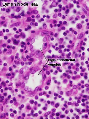 | 300 × 400 (52 KB) | S8600021 (talk | contribs) | ==Lymph Node Histology== Original file name: Lyn42he.jpg http://www.lab.anhb.uwa.edu.au/mb140/CorePages/Lymphoid1/lymph1.htm#Lymph Lymph node histology 05.jpg {{Template:Blue Histology}} Category:Immune |
You cannot overwrite this file.