File:Lutein cell lipid and glycogen em02.jpg

Original file (1,109 × 796 pixels, file size: 227 KB, MIME type: image/jpeg)
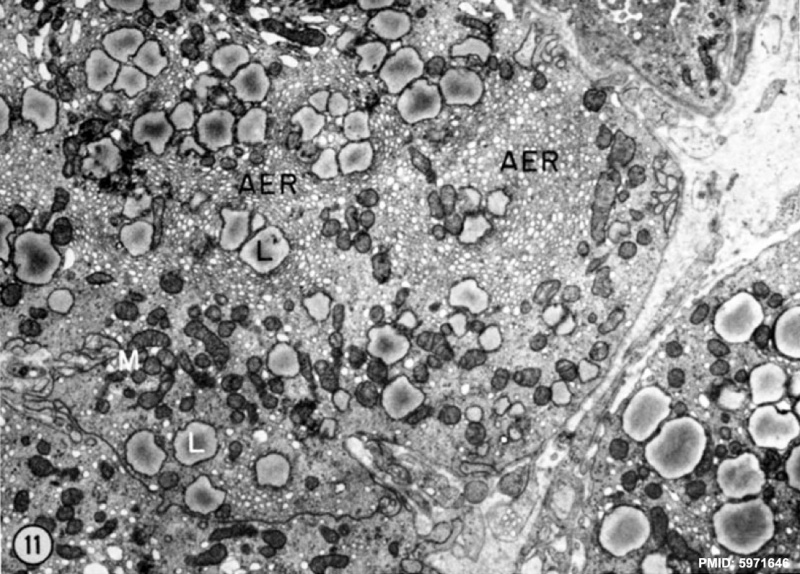
Lutein Cell Lipid and Glycogen EM
Lutein cell from a corpus luteum of pregnancy, 15 days after ovulation. Agranular endoplasmic reticulum (AER) is organized in circular aggregations of vesicles as well as short, branched tubules surrounding mitochondria (M) and lipid droplets (L), which average 1.0 um in size and show little electron opacity. This tissue was fixed with a hypo-osmotic (~50 milliosmols) solution of osmium tctroxide. The dilation of the vesicular profiles of the agranular endoplasmic reticulum is apparent. X 7,000.
- Agranular endoplasmic reticulum (AER) is the historic name for what is now know as Smooth Endoplasmic Reticulum (SER). SER is abundant in these cells as it synthesizes lipids, phospholipids and steroids.
Reference
<pubmed>5971646</pubmed>
Copyright
Rockefeller University Press - Copyright Policy This article is distributed under the terms of an Attribution–Noncommercial–Share Alike–No Mirror Sites license for the first six months after the publication date (see http://www.jcb.org/misc/terms.shtml). After six months it is available under a Creative Commons License (Attribution–Noncommercial–Share Alike 4.0 Unported license, as described at https://creativecommons.org/licenses/by-nc-sa/4.0/ ). (More? Help:Copyright Tutorial)
Ovarian steroid cells. II. The lutein cell
J Cell Biol. 1966 Dec;31(3):517-42.
Blanchette EJ.
Abstract
The lutein cells of the rabbit exhibit fine structural variations during their life-span of 28 to 30 days. A systematic examination of the corpus luteum reveals that cellular distinctions may be recognized during the first, second, and third stages of pregnancy. The agranular endoplasmic reticulum reveals vesicular, tubular, and cisternal profiles after fixation with each of the following fixatives: glutaraldehyde, osmium tetroxide, and permanganate. The osmolality of the fixing solutions was varied with sucrose and recorded with an osmometer in order to determine the effect of osmotic concentration on the intracellular membranous profiles. It was determined that vesicles and short, branched tubules of similar structure are present in the agranular reticulum when the osmolalities are 300 to 800 milliosmols (iso-osmotic considered 300 milliosmols). At 900 milliosmols, the vesicular or tubular lumen is obliterated. Intracellular membrane profiles do not exhibit interconversions due to hyperosmotic fixative solutions. The agranular endoplasmic reticulum is randomly distributed as short tubular profiles during the first third of pregnancy. A continuity between these membranes and irregular, electron-opaque lipid masses is evident. When physiological and histochemical data indicate that the lutein cell may be storing sterol precursors, cytological observations show that the agranular endoplasmic reticulum exists in a more organized pattern within the cytoplasmic matrix. Vesicular and short tubular, circular aggregations as well as whorled cisternal patterns surround the larger, less electron-opaque lipid droplets. Surface views of cisternal agranular endoplasmic reticulum exhibit tubular extensions, accentuating the continuity between these two profiles. During the progress of pregnancy, the lutein cell increases in diameter, and accumulates both lipid inclusions and aggregations of intracellular membranes. The agranular endoplasmic reticulum may be peripherally packed and arranged parallel to the cell surface during later stages. In the postpartum, degenerating lutein cell, large myelin figures are present which form from the agranular endoplasmic reticulum. These cellular events are discussed in relation to lutein cell activity, including both secretion of product and storage of precursors.
PMID 5971646
File history
Click on a date/time to view the file as it appeared at that time.
| Date/Time | Thumbnail | Dimensions | User | Comment | |
|---|---|---|---|---|---|
| current | 01:30, 14 February 2013 | | 1,109 × 796 (227 KB) | Z8600021 (talk | contribs) | |
| 01:24, 14 February 2013 | 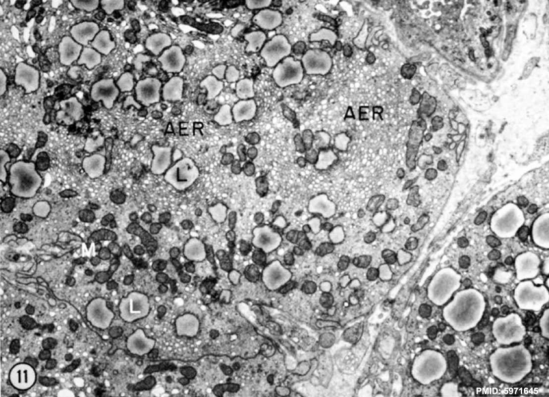 | 1,108 × 802 (230 KB) | Z8600021 (talk | contribs) | ==Lutein Cell lipid and Glycogen EM== ==Reference== <pubmed>5971645</pubmed> {{JCB}} ---- ===Ovarian steroid cells. I. Differentiation of the lutein cell from the granulosa follicle cell during the preovulatory stage and under the influence of exo |
You cannot overwrite this file.
File usage
There are no pages that use this file.