File:Lutein cell glycogen granule em01.jpg

Original file (1,149 × 749 pixels, file size: 169 KB, MIME type: image/jpeg)
Lutein Cell Glycogen Granule EM
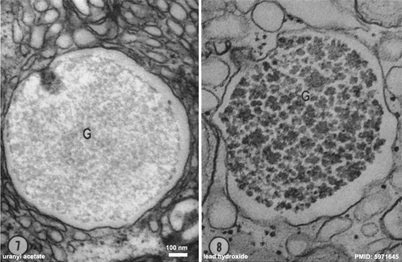
- Figure 7 - A 1 um membrane-bounded vacuole containing glycogen (G) within a lutein cell during the first third of pregnancy. Stained with uranyl acetate. X 54,000. Alpha glycogen particles are not stained by uranyl acetate.
- Figure 8 - A membrane-bounded vacuole containing glycogen (G) in a lutein cell, similar to that of Fig. 7. Stained with lead hydroxide. X 54,000. Alpha glycogen particles exhibit a deep staining quality with lead saIts.
Reference
<pubmed>5971645</pubmed>
Copyright
Rockefeller University Press - Copyright Policy This article is distributed under the terms of an Attribution–Noncommercial–Share Alike–No Mirror Sites license for the first six months after the publication date (see http://www.jcb.org/misc/terms.shtml). After six months it is available under a Creative Commons License (Attribution–Noncommercial–Share Alike 4.0 Unported license, as described at https://creativecommons.org/licenses/by-nc-sa/4.0/ ). (More? Help:Copyright Tutorial)
Ovarian steroid cells. I. Differentiation of the lutein cell from the granulosa follicle cell during the preovulatory stage and under the influence of exogenous gonadotrophins
J Cell Biol. 1966 Dec;31(3):501-16.
Blanchette EJ.
Abstract
The granulosa follicle cell of the Graafian follicle of the rabbit ovary differentiates into a lutein cell involved in steroid synthesis. Cytological events which occur within the granulosa cell of the normally stimulated follicle prior to ovulation have been duplicated by the intrafollicular injection of exogenous gonadotrophin. The luteinization of the granulosa cells involves the accumulation of 250- to 300-A, electron-opaque, spherical granules, dispersed within the cytoplasmic matrix, which have been identified as glycogen with the PAS-staining procedure. Further development of the granulosa cell following ovulation involves an increase in cell size, a decrease in the number of RNP particles, and an accumulation of an abundant system of intracellular membranes (agranular endoplasmic reticulum). Glycogen granules first appear in the granulosa cells as the separate, monoparticulate form. After follicle rupture and the formation of agranular endoplasmic reticulum, glycogen particles are present in a rosette arrangement within membrane-bounded vacuoles. The rosette arrangement of glycogen particles is also found dispersed within the cytoplasmic matrix of the lutein cell during the later stages of the cell life-span. Injection of luteinizing hormone or human chorionic gonadotrophin into a mature follicle also produces a marked accumulation of monoparticulate glycogen in the majority of granulosa cells, within 30 min. Cytoplasmic extensions which contain the glycogen masses are noticeably free of RNP particles.
PMID 5971645
File history
Click on a date/time to view the file as it appeared at that time.
| Date/Time | Thumbnail | Dimensions | User | Comment | |
|---|---|---|---|---|---|
| current | 01:03, 14 February 2013 |  | 1,149 × 749 (169 KB) | Z8600021 (talk | contribs) | ==Lutein Cell Glycogen Granule EM== ==Reference== <pubmed>5971645</pubmed> {{JCB}} ===Ovarian steroid cells. I. Differentiation of the lutein cell from the granulosa follicle cell during the preovulatory stage and under the influence of exogenous gon |
You cannot overwrite this file.
File usage
There are no pages that use this file.