File:Lung historical image.PNG
From Embryology
Size of this preview: 800 × 441 pixels. Other resolution: 856 × 472 pixels.
Original file (856 × 472 pixels, file size: 326 KB, MIME type: image/png)
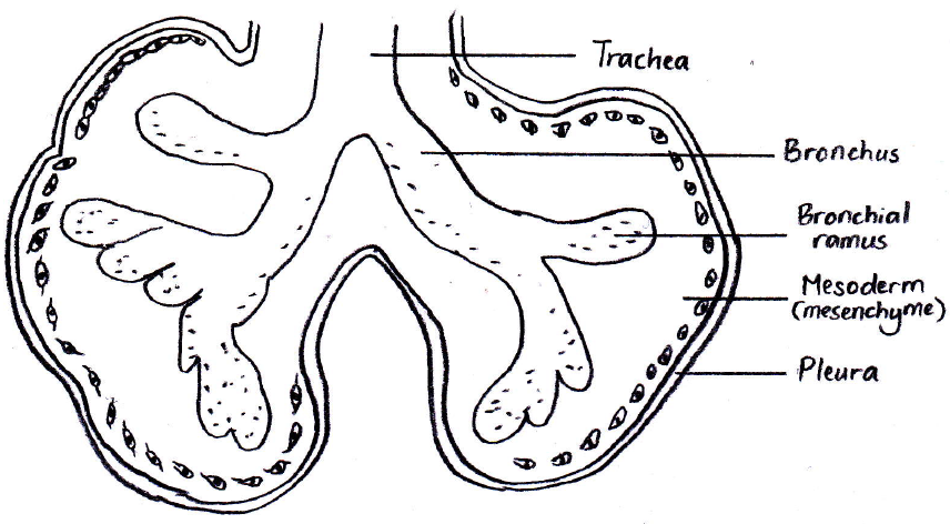
Drawing based on the diagram found in the 1921 'Text-Book of Embryology' by Bailey and Miller. It shows the rudimentary basis of lung development in the embryo, which further branching and mesenchymal differentation will for the alveoli in the fetus.
--Mark Hill (talk) 14:50 7 November 2014 (EST) Assessment - Student drawn figure relates to project topic contains only reference. I have to ask why bother redrawing a figure that already exists on the website? You have not added any additional information related to fetal development.
File history
Click on a date/time to view the file as it appeared at that time.
| Date/Time | Thumbnail | Dimensions | User | Comment | |
|---|---|---|---|---|---|
| current | 12:08, 17 October 2014 | | 856 × 472 (326 KB) | Z3372817 (talk | contribs) | Hand-drawn image showing the rudimentary basis of lung development in the embryo, which with further branching and mesenchymal differentation, will form the alveoli in the fetus. Drawing based on the diagram found in the 1921 'Text-Book of Embryology'... |
| 16:40, 6 October 2014 |  | 856 × 472 (326 KB) | Z3372817 (talk | contribs) | Drawing based on the diagram found in the 1921 'Text-Book of Embryology' by Bailey and Miller. It shows the rudimentary basis of lung development in the embryo, which further branching and mesenchymal differentation will for the alveoli in the fetus. |
You cannot overwrite this file.
File usage
The following 2 pages use this file: