File:Lowsley1912 fig08.jpg

Original file (1,324 × 1,196 pixels, file size: 312 KB, MIME type: image/jpeg)
Fig. 8. New-born Baby 36 cm Prostate
x 6, camera lucida.
| Historic Disclaimer - information about historic embryology pages |
|---|
 |
- Links: fig 1 | fig 2 | fig 3 | fig 4 | fig 5 | fig 6 | fig 7 | fig 8 | table 1 | table 2 | plate 1 | plate 2 | plate 3 | Prostate Development | Lowsley 1912
- Human Fetal Prostate
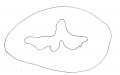
Fig 1. Fetus 5 cm
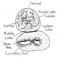
Fig. 2. Fetus 7.5 cm
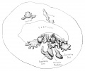
Fig. 3. Fetus 12.5 cm
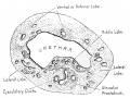
Fig. 4. Fetus 12.5 cm
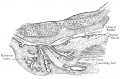
Fig. 5. Fetus 16 cm
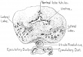
Fig. 6. Fetus 19 cm
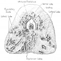
Fig. 7. Fetus 27 cm
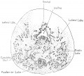
Fig. 8. New-born 36 cm
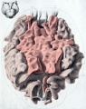
Plate 1. New-born (dorsal view)
Plate 2. New-born (sagittal view)
Plate 3. New-born (posterior and lateral lobe removed)


Reference
Lowsley OS. The development of the human prostate gland with reference to the development of other structures at the neck of the urinary bladder. (1912) Amer. J Anat. 13(3): 299-346.
Cite this page: Hill, M.A. (2024, April 19) Embryology Lowsley1912 fig08.jpg. Retrieved from https://embryology.med.unsw.edu.au/embryology/index.php/File:Lowsley1912_fig08.jpg
- © Dr Mark Hill 2024, UNSW Embryology ISBN: 978 0 7334 2609 4 - UNSW CRICOS Provider Code No. 00098G
File history
Click on a date/time to view the file as it appeared at that time.
| Date/Time | Thumbnail | Dimensions | User | Comment | |
|---|---|---|---|---|---|
| current | 21:36, 16 June 2016 | | 1,324 × 1,196 (312 KB) | Z8600021 (talk | contribs) | |
| 21:32, 16 June 2016 | 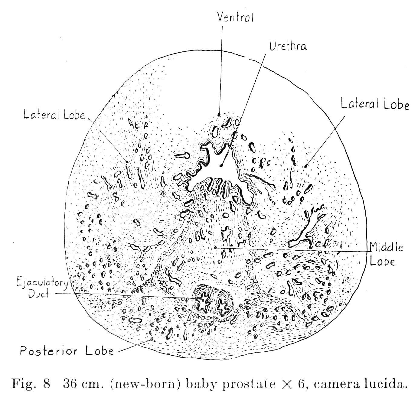 | 1,382 × 1,313 (333 KB) | Z8600021 (talk | contribs) |
You cannot overwrite this file.
File usage
The following 16 pages use this file:
- Paper - The development of the human prostate gland with reference to the development of other structures at the neck of the urinary bladder (1912)
- Prostate Development
- File:Lowsley1912 fig01.jpg
- File:Lowsley1912 fig02.jpg
- File:Lowsley1912 fig03.jpg
- File:Lowsley1912 fig04.jpg
- File:Lowsley1912 fig05.jpg
- File:Lowsley1912 fig06.jpg
- File:Lowsley1912 fig07.jpg
- File:Lowsley1912 fig08.jpg
- File:Lowsley1912 plate01.jpg
- File:Lowsley1912 plate02.jpg
- File:Lowsley1912 plate03.jpg
- File:Lowsley1912 table01.jpg
- File:Lowsley1912 table02.jpg
- Template:Lowsley1912 figures