File:Long1911 plate06.jpg

Original file (1,070 × 1,500 pixels, file size: 291 KB, MIME type: image/jpeg)
Plate 6
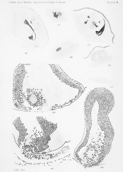
Figs. 32-37. First polar cells from oviducal eggs which contain the second spindle. They form a series of steps which illustrate the degeneration of the first polar cell. Figs. 32 and 33 are of polar cells which have divided into two or more parts. X (2500) 2000. Figs. 38-40. Three stages in the process of ovulation. In all three cases the egg contains the second spindle and is accompanied by the first polar cell. X(i7o) 136.
Note. A minute body appearing in the clear space between zona pellucida and vitellus in fig. 38 is due to a defect in the plate.
| Historic Disclaimer - information about historic embryology pages |
|---|
 |
File history
Click on a date/time to view the file as it appeared at that time.
| Date/Time | Thumbnail | Dimensions | User | Comment | |
|---|---|---|---|---|---|
| current | 20:23, 21 April 2014 | | 1,070 × 1,500 (291 KB) | Z8600021 (talk | contribs) | ==Plate 1== {{Long1911 figures}} |
You cannot overwrite this file.
File usage
The following page uses this file: