File:Long1911 plate05.jpg

Original file (1,070 × 1,500 pixels, file size: 87 KB, MIME type: image/jpeg)
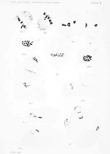
Plate 5 Second Spindle and Formation op Second Polar Cell
Figs. 24a, 246. A spindle similar to that of fig. 23, cut into two parts. There are 20 chromosomes. First polar cell absent. Oviducal egg. X(25oo) 2000.
Figs. 25-27. Old second spindles from three eggs showing diminution of circumpolar bodies. All from oviducal eggs without first polar cell. X(2 5oo) 2000.
Figs. 28a, 28b. Polar views of the two daughter plates of a dividing second spindle in a stage corresponding to that in fig. 16, plate 3. The first polar cell is very small. Oviducal egg. X (2500) 2000.
Figs. 29a, 29ft. Two sections of an oviducal egg. The oblique spindle is more advanced than the one in fig. 28. The stage of the abstriction of the polar cell (see also fig. J, p. 40) corresponds to that of figs. 16-17. The first polar cell is seen lying at the left of the second in fig. 29b. The egg contains the heads of two spermatozoa. X(25oo) 2000.
Fig. 30. Oviducal egg showing second polar cell newly abstricted, the first polar cell, and the head of a spermatozoon. X (1200) 960.
Figs. 31a, 316. Two sections of an egg (another section of which, less highly magnified, is shown in fig. 40) exhibiting in fig. 31a the first polar cell lying in the enlarged perivitelline space. X(i20o) 960.
| Historic Disclaimer - information about historic embryology pages |
|---|
| Pages where the terms "Historic" (textbooks, papers, people, recommendations) appear on this site, and sections within pages where this disclaimer appears, indicate that the content and scientific understanding are specific to the time of publication. This means that while some scientific descriptions are still accurate, the terminology and interpretation of the developmental mechanisms reflect the understanding at the time of original publication and those of the preceding periods, these terms, interpretations and recommendations may not reflect our current scientific understanding. (More? Embryology History | Historic Embryology Papers) |
File history
Click on a date/time to view the file as it appeared at that time.
| Date/Time | Thumbnail | Dimensions | User | Comment | |
|---|---|---|---|---|---|
| current | 20:22, 21 April 2014 |  | 1,070 × 1,500 (87 KB) | Z8600021 (talk | contribs) | ==Plate 5== {{Long1911 figures}} |
You cannot overwrite this file.
File usage
The following page uses this file:
