File:Lockwood1888b fig49.jpg
Lockwood1888b_fig49.jpg (800 × 496 pixels, file size: 69 KB, MIME type: image/jpeg)
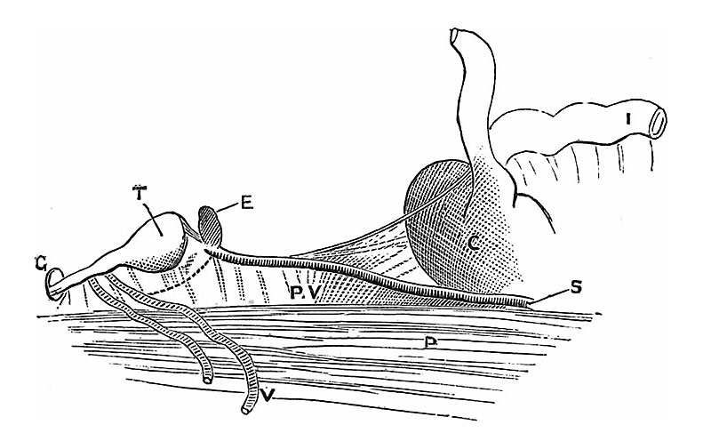
Fig. 49 Drawing made from a seven or eight months’ foetus
To show the fold (plica vascularis) which connects the testis with the cecum. T, testicles ; E, epididymis; P, psoas; V, vas deferens; G, plica gubernatrix, disappearing into processus vaginalis; P.V, plica vascularis ; C, cecum ; S,spermatic artery ; I, ilium.
Reference
Lockwood CB. Development and transition of the testis, normal and abnormal. (1888) J Anat. 22(4):505-41. PMID 17231761
Cite this page: Hill, M.A. (2024, April 17) Embryology Lockwood1888b fig49.jpg. Retrieved from https://embryology.med.unsw.edu.au/embryology/index.php/File:Lockwood1888b_fig49.jpg
- © Dr Mark Hill 2024, UNSW Embryology ISBN: 978 0 7334 2609 4 - UNSW CRICOS Provider Code No. 00098G
File history
Click on a date/time to view the file as it appeared at that time.
| Date/Time | Thumbnail | Dimensions | User | Comment | |
|---|---|---|---|---|---|
| current | 17:10, 14 April 2020 | | 800 × 496 (69 KB) | Z8600021 (talk | contribs) | adjust size W 800px |
| 17:10, 14 April 2020 |  | 927 × 575 (82 KB) | Z8600021 (talk | contribs) | crop | |
| 17:09, 14 April 2020 |  | 1,213 × 799 (136 KB) | Z8600021 (talk | contribs) |
You cannot overwrite this file.
File usage
The following page uses this file: