File:Lockwood1888a plate07.jpg
Original file (1,280 × 985 pixels, file size: 285 KB, MIME type: image/jpeg)
Plate VII
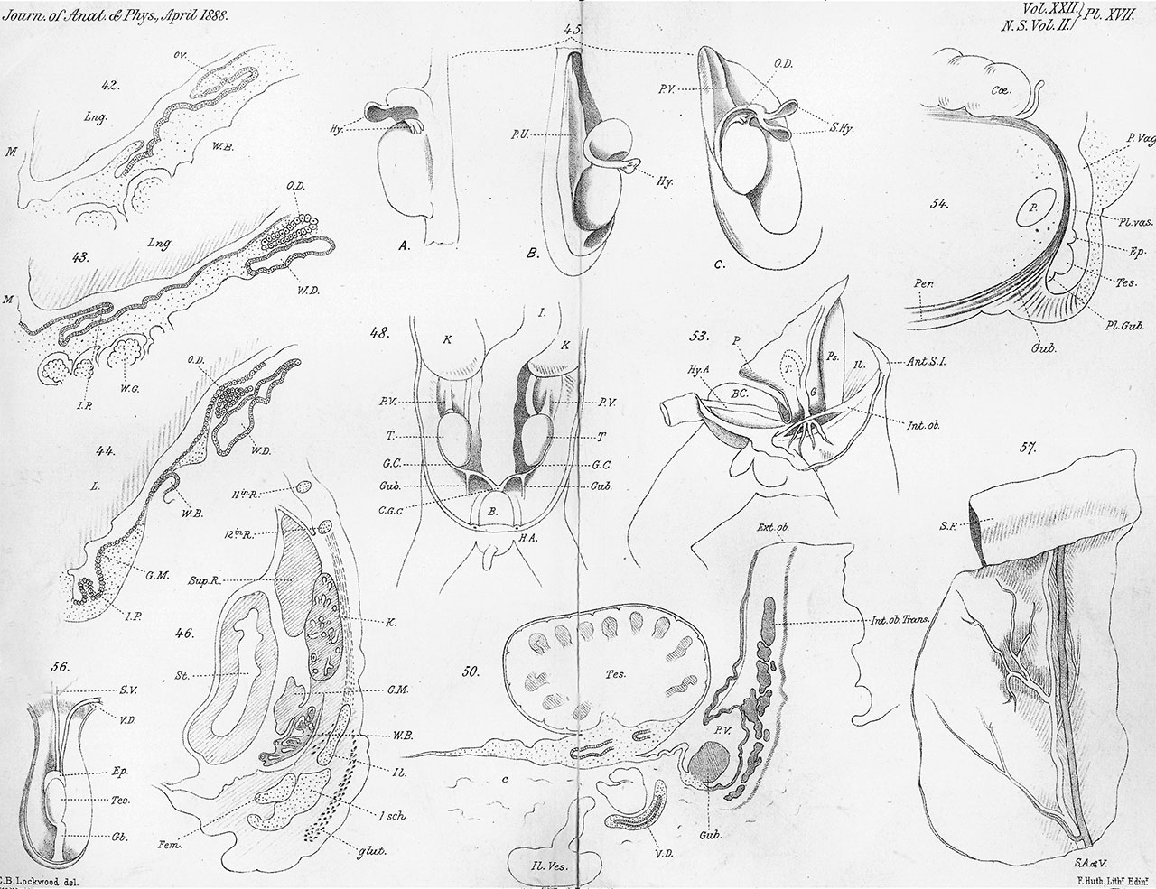
Fig. 42. Opening of oviduct of embryo pig into peritoneal cavity. Ov., oviduct ; W.B., Wolffian body ; Lng., lung; M, mesentery.
Fig. 43. Oviduct, where its lumen begins. Letters the same as fig. 42. J.P., inner process; W.G., Wolffian glomeruli.
Fig. 44. Oviduct near it hinder end. Letters the same as figs. 42 and 43. O.D., oviduct; G.I, genital mass.
Fig. 45. A, B, and C, testicles of human fetuses ; P.V., plica vascularis ; O.D., remains of oviduct ; S.Hy., spatulate hydatid. Hy., hydatid of oviduct.
Fig. 46. Pelvic and lumbar regions of a human embryo of seventh week; #., ribs llth and 12th; Sup. &., Suprarenal body; K., kidney ; G.M., genital mass; W.B., Wolffian body ; Il., ilium ; Jsch., ischium ; Fem., femur ; Glut., gluteus maximus ; St., stomach.
Fig. 47. Electrotypes from micro-photo to show continuity of Wolffian body suprarenal (page 470).
Fig. 48. Human embryo of three months to show plica gubernatrix and position of testis; x3. 7, testicles; K., kidney; £, intestine ; P.V., plica vascularis; G.C., right and left genital cords; C.G.C, common genital cords; B., bladder; Gwub., plica gubernatrix and gubernaculum.
Fig. 49. Mesorchium and its folds (p. 60 ; woodcut).
Fig. 50. Ostium of processus vaginalis at fifth month. Tes., testicle; V.D., vas deferens; Gub., gubernaculum; P.G., plica gubernatrix ; P.V., processus vaginalis ; C., loose cellular tissue ; J/. Ves., iliac vessel ; Int. Ob. and Trans., internal oblique and transversalis muscles ; Hx. Ob., external oblique.
Fig. 51. Processus vaginalis in transverse section. Letters same as fig. 50 (woodcut).
Fig. 52. Unattached gubernaculum (woodcut).
Fig. 53. Gubernaculum of six months’ foetus. The peritoneum has been dissected from psoas and iliacus, and turned over the bladder in order to show the gubernaculum upon its outer surface ; 'P., peritoneum; Ant. S.Z., anterior superior spine of ilium; B. bladder; Hy. A., hypogastric arteries; 7., testicle; .G., gubernaculum ; Ps., psoas ; J1., iliacus ; Jné. Ob., internal oblique.
Fig. 54. Diagram constructed from sections and dissections of human foetuses at full time. To show peritoneal, scrotal, and perineal prolongations of the gubernaculum testis; Ce., cecum ; P. Vag., processus vaginalis; P., pubic bone ; Pl. Vas., plica vascularis ; Ep., epididymis; Tes., testicle; Pl. Gub., plica gubernatrix; G‘ub., gubernaculum ; Per., perineal fibres of the gubernaculum testis. |
Fig. 55. Infantile hernia to show the band of muscular fibres passing from epididymis to sac of hernia (woodcut).
Fig. 56. Processus vaginalis at eighth month of intra-uterine life to show the relations of the vas deferens and spermatic vessels to its posterior wall; S.V., spermatic vessels; V.D., vas deferens ; Ep., epididymis ; Tes., testicle; Gb., gubernaculum.
Fig. 57. Recurring branches of spermatic artery; S. A. and V,, spermatic artery and vein ; S., sigmoid flexure.
Reference
Lockwood CB. Development and transition of the testis, normal and abnormal. (1888) J Anat. 22(3): 460.1-478. PMID 7231755
Cite this page: Hill, M.A. (2024, April 20) Embryology Lockwood1888a plate07.jpg. Retrieved from https://embryology.med.unsw.edu.au/embryology/index.php/File:Lockwood1888a_plate07.jpg
- © Dr Mark Hill 2024, UNSW Embryology ISBN: 978 0 7334 2609 4 - UNSW CRICOS Provider Code No. 00098G
File history
Click on a date/time to view the file as it appeared at that time.
| Date/Time | Thumbnail | Dimensions | User | Comment | |
|---|---|---|---|---|---|
| current | 13:11, 14 April 2020 | | 1,280 × 985 (285 KB) | Z8600021 (talk | contribs) | scale W 1280px |
| 13:11, 14 April 2020 | 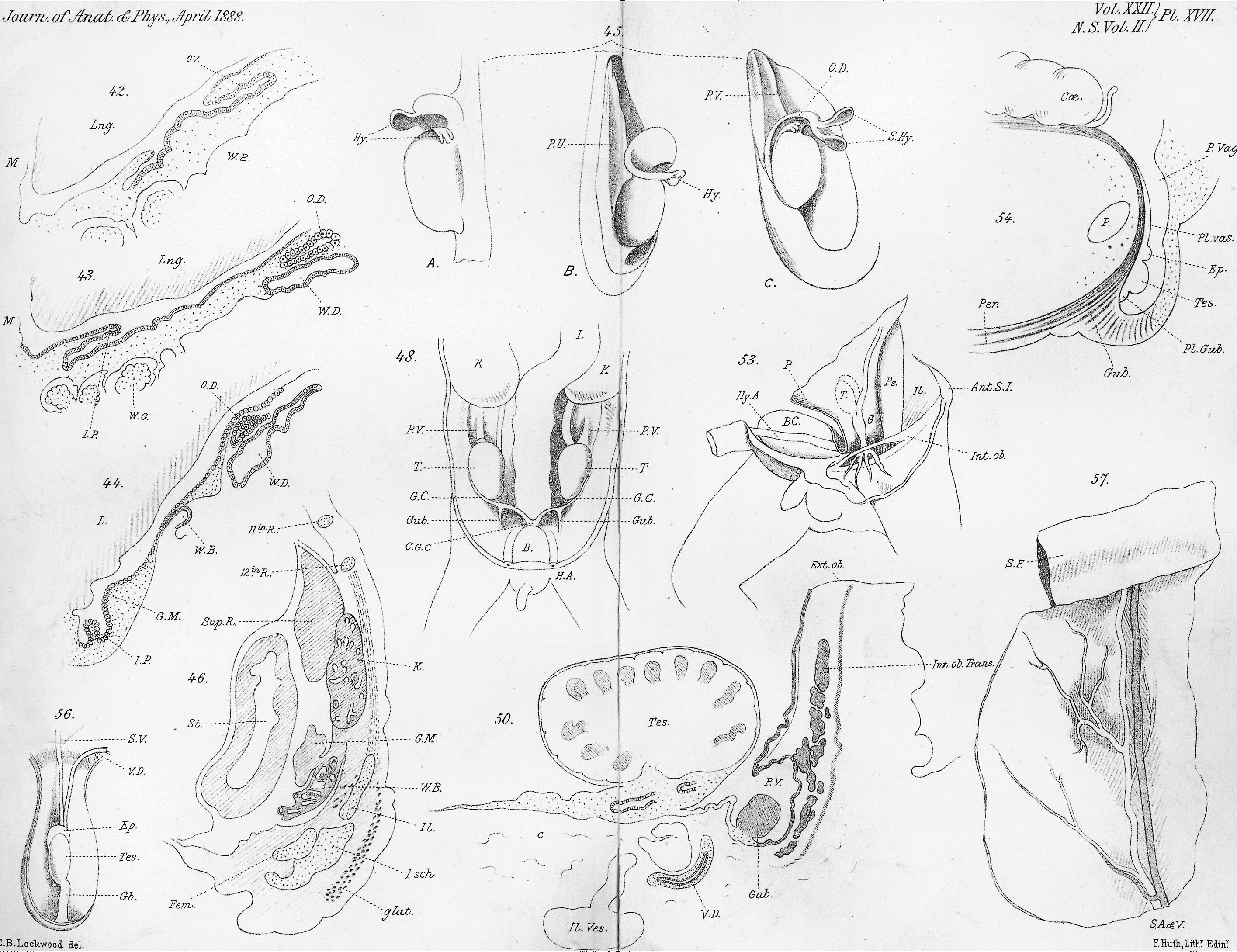 | 2,831 × 2,179 (1.14 MB) | Z8600021 (talk | contribs) | crop | |
| 13:10, 14 April 2020 | 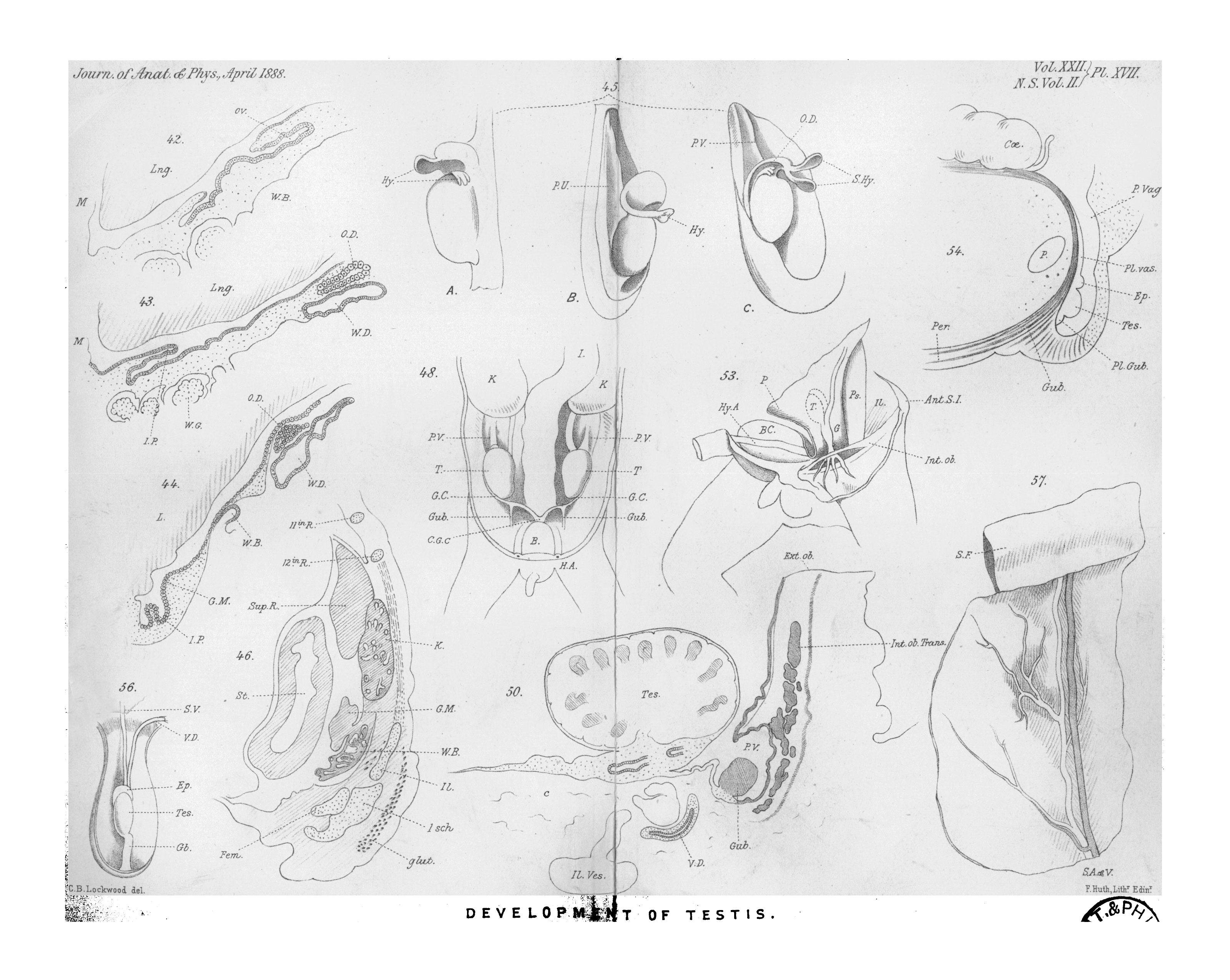 | 3,200 × 2,550 (1.03 MB) | Z8600021 (talk | contribs) | ===Reference=== {{Ref-Lockwood1888a}} {{Footer}} Category:TestisCategory:MaleCategory:Genital |
You cannot overwrite this file.
File usage
The following page uses this file: