File:Lockwood1887b fig30.jpg
From Embryology

Size of this preview: 775 × 600 pixels.
Original file (800 × 619 pixels, file size: 69 KB, MIME type: image/jpeg)
Plate II
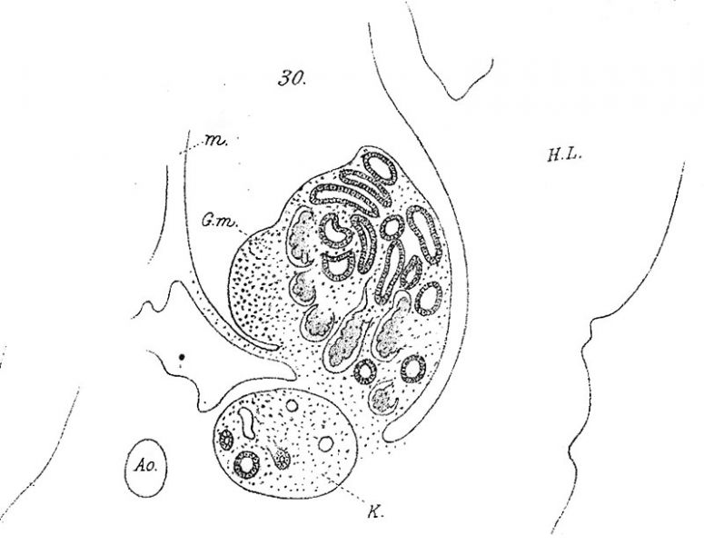
Fig. 30. Rabbit, fourteenth day, to show relation of hinder part of the Wolffian body and genital mass to one another, and to the kidney which has just appeared. GJ, genital mass; AO, aorta; XK, kidney ; HZ, hind limb; M, mesentery. x 25.
Reference
Lockwood CB. Development and transition of the testis, normal and abnormal. (1887) J Anat. 22(1): 38-77. PMID 17231729
Cite this page: Hill, M.A. (2024, April 18) Embryology Lockwood1887b fig30.jpg. Retrieved from https://embryology.med.unsw.edu.au/embryology/index.php/File:Lockwood1887b_fig30.jpg
- © Dr Mark Hill 2024, UNSW Embryology ISBN: 978 0 7334 2609 4 - UNSW CRICOS Provider Code No. 00098G
File history
Click on a date/time to view the file as it appeared at that time.
| Date/Time | Thumbnail | Dimensions | User | Comment | |
|---|---|---|---|---|---|
| current | 19:37, 14 April 2020 | | 800 × 619 (69 KB) | Z8600021 (talk | contribs) |
You cannot overwrite this file.
File usage
The following page uses this file: