File:Lineback1920 fig06-7.jpg
From Embryology

Size of this preview: 800 × 539 pixels. Other resolution: 1,200 × 808 pixels.
Original file (1,200 × 808 pixels, file size: 177 KB, MIME type: image/jpeg)
Cross-section of the Ascending Colon
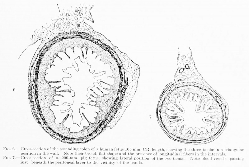
Fig. 6. Cross-section of the ascending colon of a human fetus
Cross-section of the ascending colon of a human fetus 105 mm. CR. length, showing the three tseniaj in a triangular position in the wall. Note their broad, flat shape and the presence of longitudinal fibers in the intervals.
Fig. 7 Cross-section of the ascending colon of a pig fetus
Cross-section of a 200-mm. pig fetus, showing lateral position of the two teniae. Note blood-vessels passing just beneath the peritoneal layer to the vicinity of the bands.
File history
Click on a date/time to view the file as it appeared at that time.
| Date/Time | Thumbnail | Dimensions | User | Comment | |
|---|---|---|---|---|---|
| current | 08:48, 27 December 2012 |  | 1,200 × 808 (177 KB) | Z8600021 (talk | contribs) |
You cannot overwrite this file.
File usage
The following page uses this file: