File:Licata1954 fig14.jpg

Original file (1,000 × 1,347 pixels, file size: 177 KB, MIME type: image/jpeg)
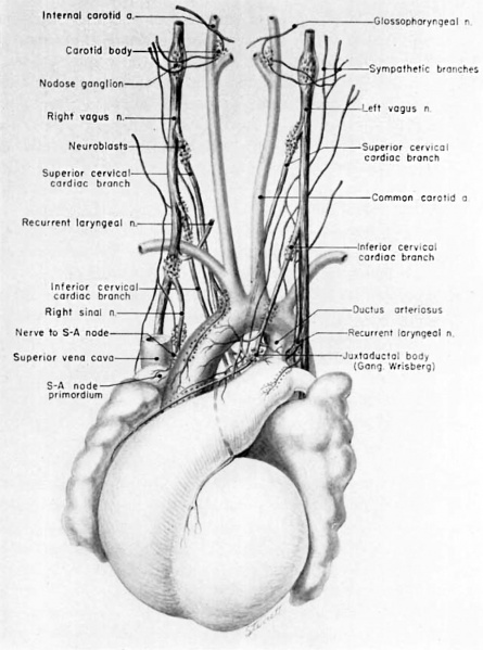
Fig. 14 Ventral aspect of heart and great vessels of a 31.5 mm embryo
Showing the distribution of the cardiac nerves and ganglia. For greater clarity, the vessels and nerves cephalic to the heart have been represented as if pulled out somewhat longer than their true proportions. The aortic arch is shown as if drawn craniad so the relations of the ductus arteriosus and the nerves could be more readily shown.
(Reconstruction X 100, illustration X 50.)
- Links: fig 1 | fig 2 | fig 3 | fig 4 | fig 5 | fig 6 | fig 7 | fig 8 | fig 9 | fig 10 | fig 11 | fig 12 | fig 13 | fig 14 | fig 15 | fig 16 | fig 16a | fig 16b | fig 16c | fig 16d | 1954 Licata | Historic Papers | Heart Development
Reference
Licata RH. The human embryonic heart in the ninth week. (1954) Amer. J Anat., 94: 73-125. PMID 13124266
Cite this page: Hill, M.A. (2024, April 25) Embryology Licata1954 fig14.jpg. Retrieved from https://embryology.med.unsw.edu.au/embryology/index.php/File:Licata1954_fig14.jpg
- © Dr Mark Hill 2024, UNSW Embryology ISBN: 978 0 7334 2609 4 - UNSW CRICOS Provider Code No. 00098G
File history
Click on a date/time to view the file as it appeared at that time.
| Date/Time | Thumbnail | Dimensions | User | Comment | |
|---|---|---|---|---|---|
| current | 11:51, 5 March 2017 |  | 1,000 × 1,347 (177 KB) | Z8600021 (talk | contribs) | |
| 11:51, 5 March 2017 |  | 1,343 × 2,051 (384 KB) | Z8600021 (talk | contribs) |
You cannot overwrite this file.
File usage
The following 4 pages use this file: