File:Licata1954 fig13.jpg
From Embryology

Size of this preview: 655 × 599 pixels. Other resolution: 1,000 × 915 pixels.
Original file (1,000 × 915 pixels, file size: 114 KB, MIME type: image/jpeg)
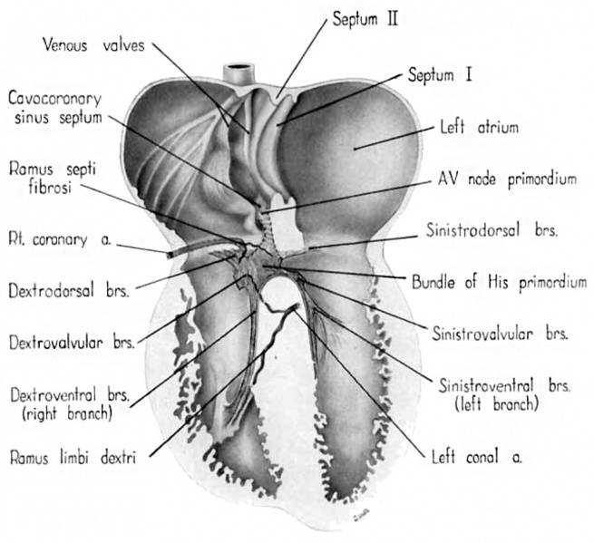
Fig. 13 Semischematic diagram to show the branches of the coronary arteries supplying the bundle of His and its main branches
The illustration was drawn as if the composite mass of endocardial cushion tissue which oceludes the interventricular foramen had been completely removed to expose the bundle of His.
- Links: fig 1 | fig 2 | fig 3 | fig 4 | fig 5 | fig 6 | fig 7 | fig 8 | fig 9 | fig 10 | fig 11 | fig 12 | fig 13 | fig 14 | fig 15 | fig 16 | fig 16a | fig 16b | fig 16c | fig 16d | 1954 Licata | Historic Papers | Heart Development
Reference
Licata RH. The human embryonic heart in the ninth week. (1954) Amer. J Anat., 94: 73-125. PMID 13124266
Cite this page: Hill, M.A. (2024, April 24) Embryology Licata1954 fig13.jpg. Retrieved from https://embryology.med.unsw.edu.au/embryology/index.php/File:Licata1954_fig13.jpg
- © Dr Mark Hill 2024, UNSW Embryology ISBN: 978 0 7334 2609 4 - UNSW CRICOS Provider Code No. 00098G
File history
Click on a date/time to view the file as it appeared at that time.
| Date/Time | Thumbnail | Dimensions | User | Comment | |
|---|---|---|---|---|---|
| current | 11:48, 5 March 2017 |  | 1,000 × 915 (114 KB) | Z8600021 (talk | contribs) | |
| 11:47, 5 March 2017 |  | 1,341 × 1,366 (251 KB) | Z8600021 (talk | contribs) |
You cannot overwrite this file.
File usage
The following 4 pages use this file: