File:LeeHalpert1932 plate02.jpg
Original file (1,241 × 1,506 pixels, file size: 314 KB, MIME type: image/jpeg)
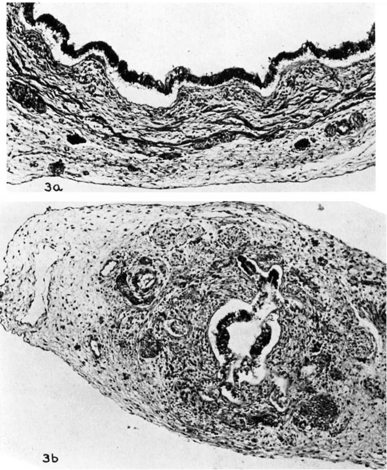
Plate 2 Fetus 130 mm Gall Bladder
3 Gall bladder of a 130 mm fetus; cross sections from the corpus (a) and from the neck (b). The surface pattern of the mucosa is rich in elevations and iudeiitatious. Two groups of deeply stained cuboidal cells situated in the periphery of the perimuseular layer are remnants of the liver primordiuni. These ‘true Luschka ducts’ make their first appearance at this stage of development (a). Some of the acinar outpocketings of the lumen of the neck appear external to the muscular coat and are lined by low columnar cells stained lightly and having their nuclei in the base of the cells (12). Photomicrographs, X 100.
Online Editor - Luschka ducts (duct of Luschka) is historic term referring to an accessory bile duct.
- Links: gallbladder
Reference
Halpert B. and Lee H. The gall bladder and the extrahepatic biliary passages in late embryonic and early fetal life. (1932) Anat. Rec. 54(1): 29-42.
Cite this page: Hill, M.A. (2024, April 25) Embryology LeeHalpert1932 plate02.jpg. Retrieved from https://embryology.med.unsw.edu.au/embryology/index.php/File:LeeHalpert1932_plate02.jpg
- © Dr Mark Hill 2024, UNSW Embryology ISBN: 978 0 7334 2609 4 - UNSW CRICOS Provider Code No. 00098G
File history
Click on a date/time to view the file as it appeared at that time.
| Date/Time | Thumbnail | Dimensions | User | Comment | |
|---|---|---|---|---|---|
| current | 15:38, 29 August 2017 | | 1,241 × 1,506 (314 KB) | Z8600021 (talk | contribs) | |
| 15:26, 29 August 2017 |  | 1,358 × 2,198 (264 KB) | Z8600021 (talk | contribs) | ===Reference=== {{Ref-LeeHalpert1932}} {{Footer}} Category:Gall BladderCategory:1930's |
You cannot overwrite this file.
File usage
There are no pages that use this file.