File:LattaTollman1937 fig11.jpg

Original file (527 × 699 pixels, file size: 87 KB, MIME type: image/jpeg)
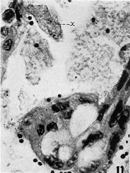
Fig. 11. Syncytial trophoblastic masses projecting into the intervillous space
In the lower mass the nuclei are shrunken and pycnotic and large vacuoles are present. Projecting from the mass at the upper right is a non-nucleated. fragment (x) containing numerous deeply staining granules. Much of the debris in the intervillous space appears as disintegrating syncytial tisue. X 600.
- Links: fig 1 | fig 2 | fig 3 | fig 4 | fig 5 | fig 6 | fig 7 | fig 8 | fig 9 | fig 10 | fig 11 | fig 12 | fig 13 | fig 14 | fig 15 | plate 1 | plate 2 | plate 3 | 1937 Latta Tollman | Historic Papers
Reference
Latta JS. and Tollman JP. An early stage of human implantation. (1937) Anat. Rec. 69(4): 443-463.
Cite this page: Hill, M.A. (2024, April 24) Embryology LattaTollman1937 fig11.jpg. Retrieved from https://embryology.med.unsw.edu.au/embryology/index.php/File:LattaTollman1937_fig11.jpg
- © Dr Mark Hill 2024, UNSW Embryology ISBN: 978 0 7334 2609 4 - UNSW CRICOS Provider Code No. 00098G
File history
Click on a date/time to view the file as it appeared at that time.
| Date/Time | Thumbnail | Dimensions | User | Comment | |
|---|---|---|---|---|---|
| current | 12:42, 26 February 2017 |  | 527 × 699 (87 KB) | Z8600021 (talk | contribs) | {{LattaTollman1937 figures}} |
You cannot overwrite this file.
File usage
The following page uses this file: