File:Kramer1942 fig08.jpg
From Embryology

Size of this preview: 800 × 366 pixels. Other resolution: 1,000 × 457 pixels.
Original file (1,000 × 457 pixels, file size: 83 KB, MIME type: image/jpeg)
Fig. 8 Semi-schematic drawings to show the relations of the various parts of the septal system in lateral aspect
(Collaboration with Bradley M. Patten).
A Based on reconstructions of the hearts of embryos 8-10 mm.
B Based on reconstructions of the hearts of embryos 12-14 mm.
| Historic Disclaimer - information about historic embryology pages |
|---|
 |
- Links:
Reference
Kramer TC. The partitioning of the truncus and conus and the formation of the membranous portion of the interventricular septum in the human heart. (1942) Amer. J Anat. 71(3): 343-370.
Cite this page: Hill, M.A. (2024, April 23) Embryology Kramer1942 fig08.jpg. Retrieved from https://embryology.med.unsw.edu.au/embryology/index.php/File:Kramer1942_fig08.jpg
- © Dr Mark Hill 2024, UNSW Embryology ISBN: 978 0 7334 2609 4 - UNSW CRICOS Provider Code No. 00098G
File history
Click on a date/time to view the file as it appeared at that time.
| Date/Time | Thumbnail | Dimensions | User | Comment | |
|---|---|---|---|---|---|
| current | 11:27, 4 February 2017 | | 1,000 × 457 (83 KB) | Z8600021 (talk | contribs) | |
| 11:25, 4 February 2017 | 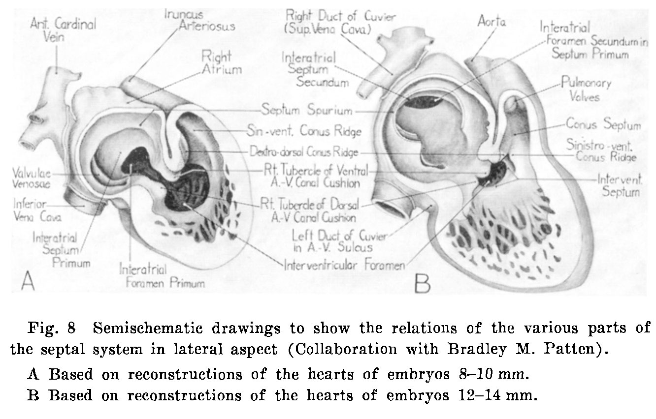 | 1,332 × 852 (193 KB) | Z8600021 (talk | contribs) | {{Kramer1942 figures}} |
You cannot overwrite this file.
File usage
The following page uses this file: