File:Kollmann741.jpg
Kollmann741.jpg (706 × 417 pixels, file size: 51 KB, MIME type: image/jpeg)
- This text is a Google translate computer generated translation and may contain many errors.
Images from - Atlas of the Development of Man (Volume 2)
(Handatlas der entwicklungsgeschichte des menschen)
- Kollmann Atlas 2: Gastrointestinal | Respiratory | Urogenital | Cardiovascular | Neural | Integumentary | Smell | Vision | Hearing | Kollmann Atlas 1 | Kollmann Atlas 2 | Julius Kollmann
- Links: Julius Kollman | Atlas Vol.1 | Atlas Vol.2 | Embryology History
| Historic Disclaimer - information about historic embryology pages |
|---|
 |
Reference
Kollmann JKE. Atlas of the Development of Man (Handatlas der entwicklungsgeschichte des menschen). (1907) Vol.1 and Vol. 2. Jena, Gustav Fischer. (1898).
Cite this page: Hill, M.A. (2024, April 23) Embryology Kollmann741.jpg. Retrieved from https://embryology.med.unsw.edu.au/embryology/index.php/File:Kollmann741.jpg
- © Dr Mark Hill 2024, UNSW Embryology ISBN: 978 0 7334 2609 4 - UNSW CRICOS Provider Code No. 00098G
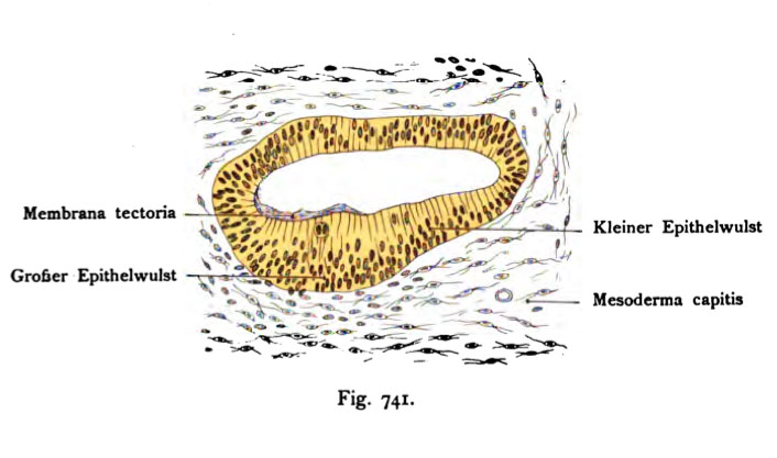
Fig. 741. Epithel des Ductus cochlearis
bei dem 2,7 cm langen Embryo eines Meerschweinchens. Unterste Windung.
(Nach Rickenbacher.)
Das Epithel zeigt den großen und kleinen Epithelwulst. Der grofee ist nochmal so dick als der kleine. Nach innen geht der große Epithelwulst ohne scharfe Grenze in das übrige Epithel über. Zwischen beiden Wülsten findet sich an der Oberfläche eine flache Einsenkung, überdies sind dort weniger Kerne vorhanden. Über den großen Epithelwulst zieht dicht anliegend ein dünnes homogenes Häutchen hinweg, das innen beginnt und nach außen sich verdünnt. Das ist das erste erkennbare Verhalten der Membrana tectoria.
File history
Click on a date/time to view the file as it appeared at that time.
| Date/Time | Thumbnail | Dimensions | User | Comment | |
|---|---|---|---|---|---|
| current | 12:30, 21 October 2011 | | 706 × 417 (51 KB) | S8600021 (talk | contribs) | {{Kollmann1907}} Category:Hearing Fig. 741. Epithel des Ductus cochlearis bei dem 2,7 cm langen Embryo eines Meerschweinchens. Unterste Windung. (Nach Rickenbacher.) Das Epithel zeigt den großen und kleinen Epithelwulst. Der grofee ist no |
You cannot overwrite this file.
File usage
The following 2 pages use this file: