File:Kollmann738.jpg
Kollmann738.jpg (505 × 503 pixels, file size: 34 KB, MIME type: image/jpeg)
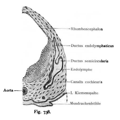
Fig. 738. Typical auditory vesicle in a human embryo of 10.2 mm CRL
(Anatomical Collection in Basel.)
Section shows the back of the head in the region of the rhombencephalon (hindbrain). Inside the auditory vesicle is a cell surface that lies beside the developing neuroepithelium medial wall, where later the utricle and sacculus will develop, in a multilayered epithelium. The same situation also occurs several times in the stratified on cochlear duct. Where no developing neuroepithelium occurs there is only a thin epithelia.
- This text is a Google translate computer generated translation and may contain many errors.
Images from - Atlas of the Development of Man (Volume 2)
(Handatlas der entwicklungsgeschichte des menschen)
- Kollmann Atlas 2: Gastrointestinal | Respiratory | Urogenital | Cardiovascular | Neural | Integumentary | Smell | Vision | Hearing | Kollmann Atlas 1 | Kollmann Atlas 2 | Julius Kollmann
- Links: Julius Kollman | Atlas Vol.1 | Atlas Vol.2 | Embryology History
| Historic Disclaimer - information about historic embryology pages |
|---|
| Pages where the terms "Historic" (textbooks, papers, people, recommendations) appear on this site, and sections within pages where this disclaimer appears, indicate that the content and scientific understanding are specific to the time of publication. This means that while some scientific descriptions are still accurate, the terminology and interpretation of the developmental mechanisms reflect the understanding at the time of original publication and those of the preceding periods, these terms, interpretations and recommendations may not reflect our current scientific understanding. (More? Embryology History | Historic Embryology Papers) |
Reference
Kollmann JKE. Atlas of the Development of Man (Handatlas der entwicklungsgeschichte des menschen). (1907) Vol.1 and Vol. 2. Jena, Gustav Fischer. (1898).
Cite this page: Hill, M.A. (2024, April 23) Embryology Kollmann738.jpg. Retrieved from https://embryology.med.unsw.edu.au/embryology/index.php/File:Kollmann738.jpg
- © Dr Mark Hill 2024, UNSW Embryology ISBN: 978 0 7334 2609 4 - UNSW CRICOS Provider Code No. 00098G
Fig. 738, Durchschnitt der Vesicula auditiva bei einem menschlichen Embryo
von 10,2 mm Nackensteifilänge.
(Anatomische Sammlung in Basel.)
Durchschnitt durch den Hinterkopf im Bereich des Rhombencephalon. Im Innern des Hörbläschens befindet sich ein Zellenbelag und zwar nimmt die Neuroepithelanlage die ganze mediale Wand ein, dort wo später der Utriculus und Sacculus sich herausbilden, kennthch an einem vielschichtigen Epithel. Dieselbe mehrfach geschichtete Lage tritt auch im Ductus cochlearis auf. An den Stellen, wo keine Neuroepithelien auftreten, ist die Lage der Epithelien dünn.
File history
Click on a date/time to view the file as it appeared at that time.
| Date/Time | Thumbnail | Dimensions | User | Comment | |
|---|---|---|---|---|---|
| current | 12:28, 21 October 2011 |  | 505 × 503 (34 KB) | S8600021 (talk | contribs) | {{Kollmann1907}} Category:Hearing Fig. 738, Durchschnitt der Vesicula auditiva bei einem menschlichen Embryo von 10,2 mm Nackensteifilänge. (Anatomische Sammlung in Basel.) Durchschnitt durch den Hinterkopf im Bereich des Rhombencephalon. |
You cannot overwrite this file.
File usage
The following 3 pages use this file:

