File:Kollmann736-737.jpg
Original file (826 × 467 pixels, file size: 56 KB, MIME type: image/jpeg)
- This text is a Google translate computer generated translation and may contain many errors.
Images from - Atlas of the Development of Man (Volume 2)
(Handatlas der entwicklungsgeschichte des menschen)
- Kollmann Atlas 2: Gastrointestinal | Respiratory | Urogenital | Cardiovascular | Neural | Integumentary | Smell | Vision | Hearing | Kollmann Atlas 1 | Kollmann Atlas 2 | Julius Kollmann
- Links: Julius Kollman | Atlas Vol.1 | Atlas Vol.2 | Embryology History
| Historic Disclaimer - information about historic embryology pages |
|---|
 |
Reference
Kollmann JKE. Atlas of the Development of Man (Handatlas der entwicklungsgeschichte des menschen). (1907) Vol.1 and Vol. 2. Jena, Gustav Fischer. (1898).
Cite this page: Hill, M.A. (2024, April 24) Embryology Kollmann736-737.jpg. Retrieved from https://embryology.med.unsw.edu.au/embryology/index.php/File:Kollmann736-737.jpg
- © Dr Mark Hill 2024, UNSW Embryology ISBN: 978 0 7334 2609 4 - UNSW CRICOS Provider Code No. 00098G
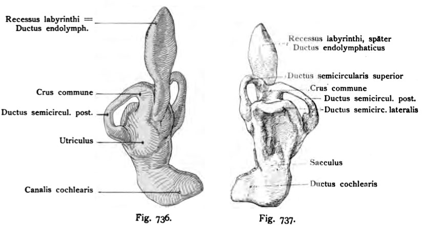
Fig. 736. Liokes Labyrinth eines mensclilichen Embryo von 13,5 Nackenlänge.
Alter etwa 5 Wochen. (Nach einer Rekonstruktion von His d. J.)
Ansicht von medial und unten. Was vor allem in dieser Figur Beachtung verdient, ist die Verbindung des Recessus labyrinthi, später Ductus endolym- phaticus, mit dem häutigen Labyrinth. Die Verbindung findet auf der Grenze zwischen der Anlage des noch vereinigten Utriculus und Sacculus statt. Nach der Trennung der beiden Säckchen bleibt der Ductus endolymphaticus mit den beiden durch einen schmalen Gang in Verbindung.
Fig. 737. Linkes Labyrinth auf einer weiteren Stufe der Differenzienins
als Fig. 735. Menschlicher Embryo von 13,5 mm Nackensteißlänge. Alter 5 Wochen.
(Nach His d. J.)
Ansicht von außen und unten. Die Einzelheiten des häutigen Labyrinths treten jetzt deutlicher hervor. Noch wenig ausgebildet sind der laterale Bogen und der Ductus cochlearis. Der letztere ist allerdings besser von dem Sacculus getrennt, als dies früher der Fall war, allein der Ductus cochlearis ist noch sehr kurz. Von dem oberen Abschnitt der Vesicula auditiva gehen die halbzirkel- förmigen Kanäle aus. Dieser Abschnitt entspricht also dem späteren Utriculus.
File history
Click on a date/time to view the file as it appeared at that time.
| Date/Time | Thumbnail | Dimensions | User | Comment | |
|---|---|---|---|---|---|
| current | 12:28, 21 October 2011 | | 826 × 467 (56 KB) | S8600021 (talk | contribs) | {{Kollmann1907}} Category:Hearing Fig. 736. Liokes Labyrinth eines mensclilichen Embryo von 13,5 Nackenlänge. Alter etwa 5 Wochen. (Nach einer Rekonstruktion von His d. J.) Ansicht von medial und unten. Was vor allem in dieser Figur Beachtung |
You cannot overwrite this file.
File usage
The following 2 pages use this file: