File:Kollmann735.jpg
Kollmann735.jpg (600 × 435 pixels, file size: 48 KB, MIME type: image/jpeg)
- This text is a Google translate computer generated translation and may contain many errors.
Images from - Atlas of the Development of Man (Volume 2)
(Handatlas der entwicklungsgeschichte des menschen)
- Kollmann Atlas 2: Gastrointestinal | Respiratory | Urogenital | Cardiovascular | Neural | Integumentary | Smell | Vision | Hearing | Kollmann Atlas 1 | Kollmann Atlas 2 | Julius Kollmann
- Links: Julius Kollman | Atlas Vol.1 | Atlas Vol.2 | Embryology History
| Historic Disclaimer - information about historic embryology pages |
|---|
 |
Reference
Kollmann JKE. Atlas of the Development of Man (Handatlas der entwicklungsgeschichte des menschen). (1907) Vol.1 and Vol. 2. Jena, Gustav Fischer. (1898).
Cite this page: Hill, M.A. (2024, April 24) Embryology Kollmann735.jpg. Retrieved from https://embryology.med.unsw.edu.au/embryology/index.php/File:Kollmann735.jpg
- © Dr Mark Hill 2024, UNSW Embryology ISBN: 978 0 7334 2609 4 - UNSW CRICOS Provider Code No. 00098G
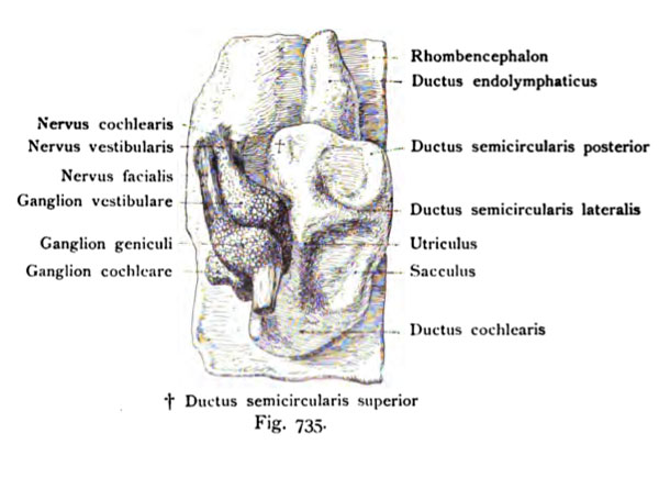
Fig. 735. Linkes Hör- oder LabyriiithbläscheiL Vesicula auditiva sioistra
mit dem Acustico facialis und seinen Ganglien. Menschlicher Embryo von 10,2 mm Nackensteißlänge. 5. Woche. 40 mal vergr. Ansicht von außen (lateral) und unten.
(Nach His d. J.)
Nach oben ragt der Recessus lab3a'inthi später Ductus endolymphaticus, nach abwärts der Anfang des Ductus cochlearis. In dem lateralen Umfang der Vesicula auditivia beginnen sich die Bogen herauszuheben. Das Ganglion cochleare und vestibuläre des Nervus acusticus liegen dicht an der Vesicula auditiva, dazwischen das Ganglion geniculi des Nervus facialis.
File history
Click on a date/time to view the file as it appeared at that time.
| Date/Time | Thumbnail | Dimensions | User | Comment | |
|---|---|---|---|---|---|
| current | 12:21, 21 October 2011 | | 600 × 435 (48 KB) | S8600021 (talk | contribs) | {{Kollmann1907}} Category:Hearing Fig. 735. Linkes Hör- oder LabyriiithbläscheiL Vesicula auditiva sioistra mit dem Acustico facialis und seinen Ganglien. Menschlicher Embryo von 10,2 mm Nackensteißlänge. 5. Woche. 40 mal vergr. Ansicht von |
You cannot overwrite this file.
File usage
The following 2 pages use this file: