File:Kollmann690.jpg

Original file (823 × 488 pixels, file size: 77 KB, MIME type: image/jpeg)
- This text is a Google translate computer generated translation and may contain many errors.
Images from - Atlas of the Development of Man (Volume 2)
(Handatlas der entwicklungsgeschichte des menschen)
- Kollmann Atlas 2: Gastrointestinal | Respiratory | Urogenital | Cardiovascular | Neural | Integumentary | Smell | Vision | Hearing | Kollmann Atlas 1 | Kollmann Atlas 2 | Julius Kollmann
- Links: Julius Kollman | Atlas Vol.1 | Atlas Vol.2 | Embryology History
| Historic Disclaimer - information about historic embryology pages |
|---|
 |
Reference
Kollmann JKE. Atlas of the Development of Man (Handatlas der entwicklungsgeschichte des menschen). (1907) Vol.1 and Vol. 2. Jena, Gustav Fischer. (1898).
Cite this page: Hill, M.A. (2024, April 25) Embryology Kollmann690.jpg. Retrieved from https://embryology.med.unsw.edu.au/embryology/index.php/File:Kollmann690.jpg
- © Dr Mark Hill 2024, UNSW Embryology ISBN: 978 0 7334 2609 4 - UNSW CRICOS Provider Code No. 00098G
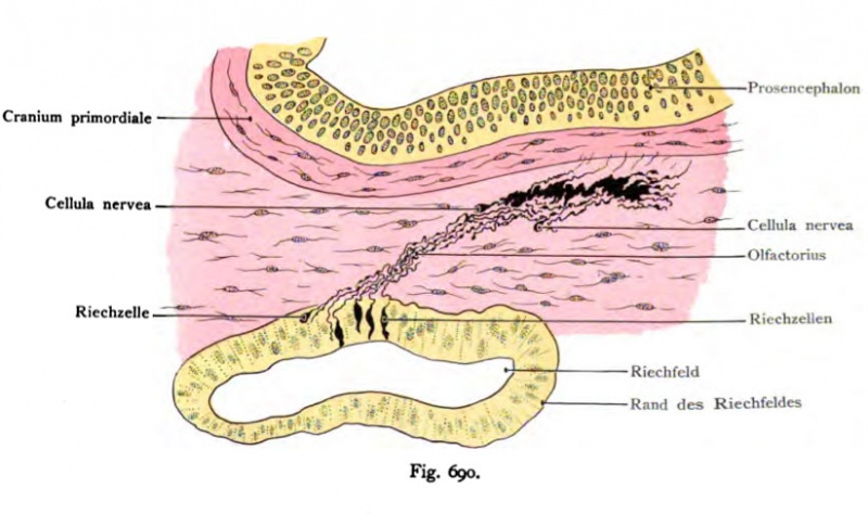
Fig. 690. Entwiclcluns des Riechnerven aus dem Epithel der Riechgrube
bei einem Hühnerembryo vom Anfang des 5. Tages. Golgipräparat.
(Nach Disse.)
Die „Riechzellen" liegen im Epithel des Riechfeldes, sie haben Spindel- oder auch Kugelform. Die spindelförmigen Zellen besitzen zwei Fortsätze, die kugeligen Zellen, die jüngsten, nur einen. Die spindelförmigen entsenden einen kurzen peripheren Fortsatz nach dem Lumen des Riechfeldes hin, den langen zentralen Fortsatz dagegen durch das Kopfmesoderm hindurch nach dem Vorderhirn. Die kugeligen Zellen werden als eine Anfangsform der Riechzellen aufgefaßt und als Cellulae nerveae bezeichnet. Am 8. Tage erreicht der Riech- nerv den Riechlappen des Gehirns und wächst mit seinen Fasern in ihn hinein. Dies ist dem Tatbestand vorauseilend in der Abbildung erkennbar.
File history
Click on a date/time to view the file as it appeared at that time.
| Date/Time | Thumbnail | Dimensions | User | Comment | |
|---|---|---|---|---|---|
| current | 10:13, 21 October 2011 | | 823 × 488 (77 KB) | S8600021 (talk | contribs) | {{Kollmann1907}} Category:Smell Fig. 690. Entwiclcluns des Riechnerven aus dem Epithel der Riechgrube bei einem Hühnerembryo vom Anfang des 5. Tages. Golgipräparat. (Nach Disse.) Die „Riechzellen" liegen im Epithel des Riechfeldes, sie h |
You cannot overwrite this file.
File usage
The following page uses this file: