File:Kollmann648.jpg

Original file (810 × 1,000 pixels, file size: 158 KB, MIME type: image/jpeg)
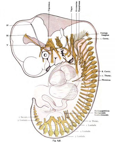
Fig. 648. Nervous system human embryo of 10.2 mm
Seen from the left side. Magnification relative to the original about 20 times.
(After His.)
The nerves are generally drawn as far as they were available, where this is not the case, indicated a cut surface. The dorsal rami of Spinal nerves are not drawn in order not to complicate too much the picture. Here too impressed by the metameric arrangement of the peripheral nervous system. The cranial nerves except the abducens with roman num- records, the spinal nerves with Arabic. The position of the extremities by dotted circle indicated by solid lines: atrial, chamber, liver and intestinal tube. The Wurzelplexus are emerging. Superior cervical plexus, inferior and lumbosacral connected by Sow. The N. phrenic, the Septum transversum reached.
- This text is a Google translate computer generated translation and may contain many errors.
Images from - Atlas of the Development of Man (Volume 2)
(Handatlas der entwicklungsgeschichte des menschen)
- Kollmann Atlas 2: Gastrointestinal | Respiratory | Urogenital | Cardiovascular | Neural | Integumentary | Smell | Vision | Hearing | Kollmann Atlas 1 | Kollmann Atlas 2 | Julius Kollmann
- Links: Julius Kollman | Atlas Vol.1 | Atlas Vol.2 | Embryology History
| Historic Disclaimer - information about historic embryology pages |
|---|
| Pages where the terms "Historic" (textbooks, papers, people, recommendations) appear on this site, and sections within pages where this disclaimer appears, indicate that the content and scientific understanding are specific to the time of publication. This means that while some scientific descriptions are still accurate, the terminology and interpretation of the developmental mechanisms reflect the understanding at the time of original publication and those of the preceding periods, these terms, interpretations and recommendations may not reflect our current scientific understanding. (More? Embryology History | Historic Embryology Papers) |
Reference
Kollmann JKE. Atlas of the Development of Man (Handatlas der entwicklungsgeschichte des menschen). (1907) Vol.1 and Vol. 2. Jena, Gustav Fischer. (1898).
Cite this page: Hill, M.A. (2024, April 25) Embryology Kollmann648.jpg. Retrieved from https://embryology.med.unsw.edu.au/embryology/index.php/File:Kollmann648.jpg
- © Dr Mark Hill 2024, UNSW Embryology ISBN: 978 0 7334 2609 4 - UNSW CRICOS Provider Code No. 00098G
Fig. 648. Nerveosystem eines meoschlicheo Embryo von 10,2 mm.
Von der linken Seite gesehen. Vergrößerung auf das Original bezogen etwa 20 mal.
(Nach His.)
Die Nerven sind im allgemeinen soweit gezeichnet, als sie vorhanden waren, wo dies nicht der Fall, ist eine Schnittfläche angedeutet. Die Rami dorsales der Spinalnerven sind nicht gezeichnet, um das Bild nicht allzusehr zu komplizieren. Auch hier imponiert die metamere Anordnung des peripheren Nervensystems. Die Hirnnerven sind mit Ausnahme des Abducens mit römischen Zahlen be- zeichnet, die Spinalnerven mit arabischen. Die Lage der Extremitäten ist durch punktierte Kreise angedeutet, durch ausgezogene Linien : Vorhof, Kammer, Leber und Darmrohr. Die Wurzelplexus sind im Entstehen. Plexus cervicalis superior, inferior und lumbosacralis durch Ansäe verbunden. Der N. phrenicus hat das Septum transversum erreicht.
File history
Click on a date/time to view the file as it appeared at that time.
| Date/Time | Thumbnail | Dimensions | User | Comment | |
|---|---|---|---|---|---|
| current | 09:44, 21 October 2011 |  | 810 × 1,000 (158 KB) | S8600021 (talk | contribs) | {{Kollmann1907}} Category:Neural Category:Spinal cord |
You cannot overwrite this file.
File usage
The following page uses this file:
