File:Kollmann646.jpg

Original file (1,000 × 606 pixels, file size: 122 KB, MIME type: image/jpeg)
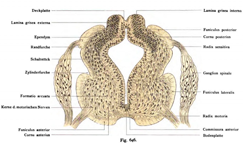
Fig. 646. The spinal cord of a human embryo of 13 mm, 5 weeks
Cross-section. (Anatomical Collection in Basel)
The Medullarkanal has a strong * bellied form of a broad Ependymschichte limited. In the ventral half of the MeduUarrohres the base plate greatly enlarged compared to the embryo of 10.2 mm in length. Your gray matter is the primary anterior cornu is, from which the Commis- sura anterior alba et grisea shows also the front and the lateral columns, the Formatis Arcuate (vord. section) and all nuclei of the motor nerves. Dorsal wing plate is in the area of the posterior horn already evident for the most part covered by the primitive dorsal column. between the Cornu is primitive anterior and posterior to the switching unit. it provides the cervix cornu posterioris, the dorsal motor nucleus (Clarkii), and the formation reticularis. Later appear in the area of the contact piece of cord cerebrospinal lateralis (= pyramidalis lateralis) (pyramidal tract side) and the fasciculus cerebello-spinalis (direct cerebellar tract).
- This text is a Google translate computer generated translation and may contain many errors.
Images from - Atlas of the Development of Man (Volume 2)
(Handatlas der entwicklungsgeschichte des menschen)
- Kollmann Atlas 2: Gastrointestinal | Respiratory | Urogenital | Cardiovascular | Neural | Integumentary | Smell | Vision | Hearing | Kollmann Atlas 1 | Kollmann Atlas 2 | Julius Kollmann
- Links: Julius Kollman | Atlas Vol.1 | Atlas Vol.2 | Embryology History
| Historic Disclaimer - information about historic embryology pages |
|---|
| Pages where the terms "Historic" (textbooks, papers, people, recommendations) appear on this site, and sections within pages where this disclaimer appears, indicate that the content and scientific understanding are specific to the time of publication. This means that while some scientific descriptions are still accurate, the terminology and interpretation of the developmental mechanisms reflect the understanding at the time of original publication and those of the preceding periods, these terms, interpretations and recommendations may not reflect our current scientific understanding. (More? Embryology History | Historic Embryology Papers) |
Reference
Kollmann JKE. Atlas of the Development of Man (Handatlas der entwicklungsgeschichte des menschen). (1907) Vol.1 and Vol. 2. Jena, Gustav Fischer. (1898).
Cite this page: Hill, M.A. (2024, April 25) Embryology Kollmann646.jpg. Retrieved from https://embryology.med.unsw.edu.au/embryology/index.php/File:Kollmann646.jpg
- © Dr Mark Hill 2024, UNSW Embryology ISBN: 978 0 7334 2609 4 - UNSW CRICOS Provider Code No. 00098G
Fig. 646. Das Rückenmark eines menschlichen Embryo von 13 mm,
5 V« Wochen alt. Querschnitt. (Anatomische Sammlung in Basel)
Der Medullarkanal besitzt eine stark gebauchte* Form, von einer breiten Ependymschichte begrenzt. In der ventralen Hälfte des MeduUarrohres hat sich die Grundplatte stark vergrößert im Vergleich zu dem Embryo von 10,2 mm Länge. Ihre graue Masse stellt das primäre Cornu anterius dar, aus dem die Commis- sura anterior grisea et alba hervorgeht, ferner die Vorder- und die Seitenstränge, die Formatis arcuata (vord. Abschnitt) und alle Kerne für die motorischen Nerven. Dorsal ist im Bereich der Flügelplatte schon das Cornu posterius erkennbar zum größten Teile bedeckt von dem primitiven Hinterstrang. Zwischen dem primitiven Cornu anterius und posterius befindet sich das Schaltstück. Es liefert das Collum cornu posterioris, den Nucleus dorsalis (Clarkii), und die Formatio reticularis. Später erscheint in dem Bereich des Schaltstückes der Fasciculus cerebrospinalis lateralis (= pyramidalis lateralis) (Pyramiden-Seitenstrangbahn) und der Fasciculus cerebello-spinalis (Kleinhirnseitenstrangbahn).
File history
Click on a date/time to view the file as it appeared at that time.
| Date/Time | Thumbnail | Dimensions | User | Comment | |
|---|---|---|---|---|---|
| current | 09:40, 21 October 2011 |  | 1,000 × 606 (122 KB) | S8600021 (talk | contribs) | {{Kollmann1907}} Category:Neural |
You cannot overwrite this file.
File usage
The following page uses this file:
