File:Kollmann645.jpg
Kollmann645.jpg (667 × 455 pixels, file size: 37 KB, MIME type: image/jpeg)
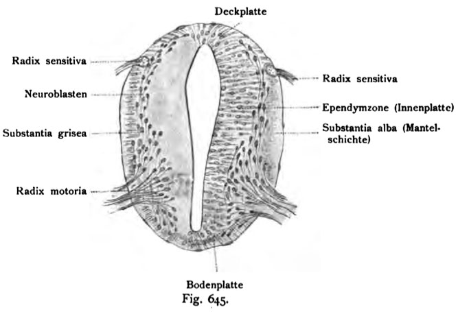
Fig. 645. Cross-section of the medulla of a four spinalis week 4 embryo
(Anatomical Collection in Basel.)
The increase of the gray and white matter with neuroblasts and Spongioblasts happens first at the two lateral halves. On the ependymal
zone, with their gray matter spongioblasts is overlaid with neuroblastoma cells
more laterally and is followed by the formation of white strands, which are in their totalitarianism
TY called the mantle. A ring of gray matter covering the ependyma and
this ring of the gray matter of the above-mentioned white "ManteP. where the
Anterior horn occurs later, the neuroblasts are mostly perpendicular to the axis
the pipe, the other parallel to the surface. The sensory root
occurs near one of the top plate, the spinal ganglion ago. The MeduUar-
channel is still very large, closed at the top of a thin top plate and bottom
by a thin base plate. In her show already ßogenfasern individual.
- This text is a Google translate computer generated translation and may contain many errors.
Images from - Atlas of the Development of Man (Volume 2)
(Handatlas der entwicklungsgeschichte des menschen)
- Kollmann Atlas 2: Gastrointestinal | Respiratory | Urogenital | Cardiovascular | Neural | Integumentary | Smell | Vision | Hearing | Kollmann Atlas 1 | Kollmann Atlas 2 | Julius Kollmann
- Links: Julius Kollman | Atlas Vol.1 | Atlas Vol.2 | Embryology History
| Historic Disclaimer - information about historic embryology pages |
|---|
| Pages where the terms "Historic" (textbooks, papers, people, recommendations) appear on this site, and sections within pages where this disclaimer appears, indicate that the content and scientific understanding are specific to the time of publication. This means that while some scientific descriptions are still accurate, the terminology and interpretation of the developmental mechanisms reflect the understanding at the time of original publication and those of the preceding periods, these terms, interpretations and recommendations may not reflect our current scientific understanding. (More? Embryology History | Historic Embryology Papers) |
Reference
Kollmann JKE. Atlas of the Development of Man (Handatlas der entwicklungsgeschichte des menschen). (1907) Vol.1 and Vol. 2. Jena, Gustav Fischer. (1898).
Cite this page: Hill, M.A. (2024, April 23) Embryology Kollmann645.jpg. Retrieved from https://embryology.med.unsw.edu.au/embryology/index.php/File:Kollmann645.jpg
- © Dr Mark Hill 2024, UNSW Embryology ISBN: 978 0 7334 2609 4 - UNSW CRICOS Provider Code No. 00098G
Fig. 645. Querschnitt der Medulla spinaiis eines vierwdchentlichen Embryo.
(Anatomische Sammlung in Basel.)
Die Vermehrung der grauen und weißen Substanz mit Neuroblasten und Spongioblasten geschieht zunächst an den beiden Seitenhälften. Auf die Ependym- zone mit ihren Spongioblasten lagert sich graue Substanz mit Neuroblastenzellen und weiter lateral folgt die Ausbildung der weißen Stränge, die man in ihrer Totali- tät als Mantel bezeichnet. Ein Ring von grauer Substanz bedeckt das Ependym und diesen Ring der grauen Substanz der ebenerwähnte weiße „ManteP. Wo das Vorderhorn später auftritt, stehen die Neuroblasten zumeist senkrecht zur Achse des Rohres, die übrigen liegen parallel zur Oberfläche. Die sensible Wurzel tritt in der Nähe der Deckplatte ein, vom Ganglion spinale her. Der MeduUar- kanal ist noch sehr groß, oben geschlossen von einer dünnen Deckplatte und unten von einer dünnen Bodenplatte. In ihr zeigen sich schon einzelne ßogenfasern.
File history
Click on a date/time to view the file as it appeared at that time.
| Date/Time | Thumbnail | Dimensions | User | Comment | |
|---|---|---|---|---|---|
| current | 09:38, 21 October 2011 |  | 667 × 455 (37 KB) | S8600021 (talk | contribs) | =Fig. 645. Cross-section of the medulla of a four spinalis week 4 embryo== (Anatomical Collection in Basel.) The increase of the gray and white matter with neuroblasts and Spongioblasts happens first at the two lateral halves. On the ependymal zone, w |
You cannot overwrite this file.
File usage
The following page uses this file:

