File:Kollmann640-641.jpg

Original file (642 × 758 pixels, file size: 61 KB, MIME type: image/jpeg)
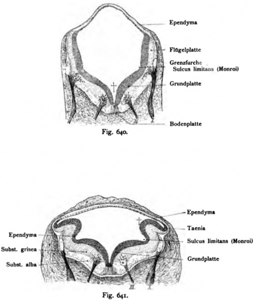
Fig. 640. Rhombencephalon (hindbrain) of a 10.2 mm long human embryo
Section 32 times larger. (Anatomical Collection in Basel.)
- This text is a Google translate computer generated translation and may contain many errors.
Images from - Atlas of the Development of Man (Volume 2)
(Handatlas der entwicklungsgeschichte des menschen)
- Kollmann Atlas 2: Gastrointestinal | Respiratory | Urogenital | Cardiovascular | Neural | Integumentary | Smell | Vision | Hearing | Kollmann Atlas 1 | Kollmann Atlas 2 | Julius Kollmann
- Links: Julius Kollman | Atlas Vol.1 | Atlas Vol.2 | Embryology History
| Historic Disclaimer - information about historic embryology pages |
|---|
| Pages where the terms "Historic" (textbooks, papers, people, recommendations) appear on this site, and sections within pages where this disclaimer appears, indicate that the content and scientific understanding are specific to the time of publication. This means that while some scientific descriptions are still accurate, the terminology and interpretation of the developmental mechanisms reflect the understanding at the time of original publication and those of the preceding periods, these terms, interpretations and recommendations may not reflect our current scientific understanding. (More? Embryology History | Historic Embryology Papers) |
Reference
Kollmann JKE. Atlas of the Development of Man (Handatlas der entwicklungsgeschichte des menschen). (1907) Vol.1 and Vol. 2. Jena, Gustav Fischer. (1898).
Cite this page: Hill, M.A. (2024, April 24) Embryology Kollmann640-641.jpg. Retrieved from https://embryology.med.unsw.edu.au/embryology/index.php/File:Kollmann640-641.jpg
- © Dr Mark Hill 2024, UNSW Embryology ISBN: 978 0 7334 2609 4 - UNSW CRICOS Provider Code No. 00098G
Fig. 640. Rhombencephalon (Rautenhirn) eines 10,2 mm langen Menschenembryo.
Schnitt. 32 mal vergrößert. (Anatomische Sammlung in Basel.)
Die mediane Furche f persistiert später als Sulcus medianus fossae rhomboideae. Die Grundplatte wird zu den Funiculi teretes. Der Sulcüs limi- tans bleibt ebenfalls erhalten; er trägt im Erwachsenen die nämliche Bezeich- nung. In der nächsten Nähe befindet sich der Ursprung des Vago-Accessorius, während ventral der motorische Hypoglossus austritt. Das Ependym bedeckt die Fossa rhomboidea in ansehnlicher Schichte, um sich schon jetzt dorsal zu einer dünnen Epithelschichte umzuändern, was später auch auf der Ober- fläche der Fossa rhomboidea geschieht.
Fig. 641. Rhombencephalon eines menschlichen Embryo von 10,2 mm Länge.
32 mal vergr. Schnitt. (Anatomische Sammlung in Basel.)
Der Ursprungskern des Vagus lateral, der des Hypoglossus medial neben dem Sulcus limitans. Die Bodenplatte und die Seitenplatte sind durch eine stark einspringende Furche: Grenzfurche, Sulcus limitans^), getrennt. Die Flügelplatte f ist stark nach außen gefaltet. Bemerkenswert ist die Umwand- lung des Ependym, das dorsal eine einfache Epithelplatte darstellt, dagegen auf der Boden-, Grund- und Flügelplatte anfangs in ansehnlicher Dicke vorkommt. Das Rhombencephalon ist umschlossen von Mesoderm, das seinerseits vom Ektoderm (einfache Linie) bedeckt ist.
File history
Click on a date/time to view the file as it appeared at that time.
| Date/Time | Thumbnail | Dimensions | User | Comment | |
|---|---|---|---|---|---|
| current | 09:29, 21 October 2011 |  | 642 × 758 (61 KB) | S8600021 (talk | contribs) | {{Kollmann1907}} Category:Neural Fig. 640. Rhombencephalon (Rautenhirn) eines 10,2 mm langen Menschenembryo. Schnitt. 32 mal vergrößert. (Anatomische Sammlung in Basel.) Die mediane Furche f persistiert später als Sulcus medianus fossae r |
You cannot overwrite this file.
File usage
The following page uses this file:
