File:Kollmann633.jpg
Kollmann633.jpg (552 × 524 pixels, file size: 39 KB, MIME type: image/jpeg)
- This text is a Google translate computer generated translation and may contain many errors.
Images from - Atlas of the Development of Man (Volume 2)
(Handatlas der entwicklungsgeschichte des menschen)
- Kollmann Atlas 2: Gastrointestinal | Respiratory | Urogenital | Cardiovascular | Neural | Integumentary | Smell | Vision | Hearing | Kollmann Atlas 1 | Kollmann Atlas 2 | Julius Kollmann
- Links: Julius Kollman | Atlas Vol.1 | Atlas Vol.2 | Embryology History
| Historic Disclaimer - information about historic embryology pages |
|---|
| Pages where the terms "Historic" (textbooks, papers, people, recommendations) appear on this site, and sections within pages where this disclaimer appears, indicate that the content and scientific understanding are specific to the time of publication. This means that while some scientific descriptions are still accurate, the terminology and interpretation of the developmental mechanisms reflect the understanding at the time of original publication and those of the preceding periods, these terms, interpretations and recommendations may not reflect our current scientific understanding. (More? Embryology History | Historic Embryology Papers) |
Reference
Kollmann JKE. Atlas of the Development of Man (Handatlas der entwicklungsgeschichte des menschen). (1907) Vol.1 and Vol. 2. Jena, Gustav Fischer. (1898).
Cite this page: Hill, M.A. (2024, April 19) Embryology Kollmann633.jpg. Retrieved from https://embryology.med.unsw.edu.au/embryology/index.php/File:Kollmann633.jpg
- © Dr Mark Hill 2024, UNSW Embryology ISBN: 978 0 7334 2609 4 - UNSW CRICOS Provider Code No. 00098G
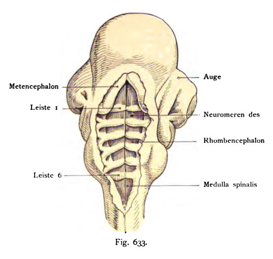
Fig. 633. Neuromeren am Myelencephalon bei Sphenodon
(einer Eidechsenart); der Kopf ist von hinten gesehen, Norma dorsalis, die Rauten- grube (Fossa rhomboidea) liegt nach Entfernung des Ektoderms und des Ependyma
ventriculi quarti vollkommen frei.
(Nach Schauinsland.)
Die Fossa rhomboidea wird durch sechs Leisten (Spinae) und mehrere symmetrische Felder geteilt, welche durch den Sulcus longitudinalis fossae rhomboideae in rechte und linke zerfallen. Die Neuromeren sind fünf an der Zahl dunkel gehalten. Die erste Leiste trennt das Metencephalon (Hinterhirn), vom Myelencephalon (Nachhirn) und die Leiste 6 des Myelencephalon von der MeduUa spinalis. Vergr. des Originales 20 mal. Die dorsalen Leisten sind gespalten und gehen sowohl dorsal als ventral ia die Seitenwand des Myelence- phalon über. Solche Neuromeren sind auch von den Säugetieren bekannt ge- worden. Die Zellen in einer Neuromere liegen anfangs dicht beisammen, und überschreiten die Grenze der folgenden Neuromere nicht.
File history
Click on a date/time to view the file as it appeared at that time.
| Date/Time | Thumbnail | Dimensions | User | Comment | |
|---|---|---|---|---|---|
| current | 17:24, 17 October 2011 |  | 552 × 524 (39 KB) | S8600021 (talk | contribs) | {{Kollmann1907}} Category:Human Category:Fetal Category:Neural Fig. 633. Neuromeren am Myelencephalon bei Sphenodon (einer Eidechsenart); der Kopf ist von hinten gesehen, Norma dorsalis, die Rauten- grube (Fossa rhomboidea) liegt nach Ent |
You cannot overwrite this file.
File usage
The following page uses this file:

