File:Kollmann623.jpg

Original file (906 × 570 pixels, file size: 46 KB, MIME type: image/jpeg)
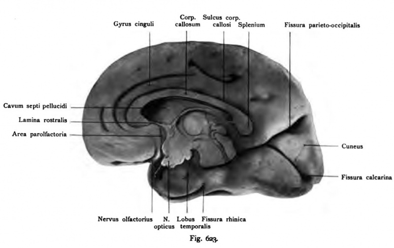
Fig. 623. The median section fetal brain from End of the 7th month
Right hemisphere. (Compare the characters 620-622 of the same brain.)
(Anatomical Collection in Basel.) Increases.
The center of the figure is occupied by the bar and the elongated cavum septum pellucidum, bounded down the front of the lamina rostralis, the rear by a continuation of the lamina terminalis (cauda) and terminalis in the middle of the lamina, which is the ceiling of the III ventricle creates the thalamus and the hypothalamus follows. From the interventricular foramen (of Monro) pulls the hypothalamic sulcus (Monro) to the rear and flows together with the cerebral aqueduct. The fissura parieto-occipitalis and the fissura cal-carina delimiting a large cuneus. The olfactory nerve is severed just above the nerve stump to find the area parolfactoria (Broca), bounded by the sulcus parolfactorius anterior and posterior. Between the area parolfactoria and the lamina terminalis and rostral extends as a small box of subcallosus gyrus (Zuckerkandl) from. At the front end of the temporal lobe, bottom area, which attracts rhinica fissure.
- This text is a Google translate computer generated translation and may contain many errors.
Images from - Atlas of the Development of Man (Volume 2)
(Handatlas der entwicklungsgeschichte des menschen)
- Kollmann Atlas 2: Gastrointestinal | Respiratory | Urogenital | Cardiovascular | Neural | Integumentary | Smell | Vision | Hearing | Kollmann Atlas 1 | Kollmann Atlas 2 | Julius Kollmann
- Links: Julius Kollman | Atlas Vol.1 | Atlas Vol.2 | Embryology History
| Historic Disclaimer - information about historic embryology pages |
|---|
| Pages where the terms "Historic" (textbooks, papers, people, recommendations) appear on this site, and sections within pages where this disclaimer appears, indicate that the content and scientific understanding are specific to the time of publication. This means that while some scientific descriptions are still accurate, the terminology and interpretation of the developmental mechanisms reflect the understanding at the time of original publication and those of the preceding periods, these terms, interpretations and recommendations may not reflect our current scientific understanding. (More? Embryology History | Historic Embryology Papers) |
Reference
Kollmann JKE. Atlas of the Development of Man (Handatlas der entwicklungsgeschichte des menschen). (1907) Vol.1 and Vol. 2. Jena, Gustav Fischer. (1898).
Cite this page: Hill, M.A. (2024, April 25) Embryology Kollmann623.jpg. Retrieved from https://embryology.med.unsw.edu.au/embryology/index.php/File:Kollmann623.jpg
- © Dr Mark Hill 2024, UNSW Embryology ISBN: 978 0 7334 2609 4 - UNSW CRICOS Provider Code No. 00098G
Fig. 623. Medianschnitt durcli das Qeliira eines menscliliclien Fetus vom
Ende des 7. Monats.
Rechte Hemisphäre. (Vergl. die Figuren 620—622 des nämlichen Gehirns.)
(Anatomische Sammlung in Basel.) Vergrößert.
Das Zentrum der Figur nimmt der Balken und das langgestreckte Cavum septi pellucidi ein, nach unten begrenzt vorn von der Lamina rostralis, hinten durch eine Fortsetzung der Lamina terminalis (Cauda) und in der Mitte von der Lamina terminalis, der sich die Decke des IIL Ventrikels anlegt Ti^er folgt der Thalamus und der Hypothalamus. Vom Foramen interventriculare (Monroi) zieht der Sulcus Hypothalamicus (Monroi) nach rückwärts und fließt mit dem Aquae- ductus cerebri zusammen. Die Fissura parieto-occipitalis und die Fissura cal- carina begrenzen einen großen Cuneus. Der Nervus olfactorius ist abgetrennt, unmittelbar oberhalb des Nervenstumpfes findet sich die Area parolfactoria (Brocae), begrenzt vom Sulcus parolfactorius anterior und posterior. Zwischen der Area parolfactoria und der Lamina rostralis und terminalis dehnt sich als kleines Feld der Gyrus subcallosus (Zuckerkandl) aus. Am vorderen Ende des Lobus temporalis, unterstes Gebiet, zieht die Fissura rhinica.
File history
Click on a date/time to view the file as it appeared at that time.
| Date/Time | Thumbnail | Dimensions | User | Comment | |
|---|---|---|---|---|---|
| current | 17:16, 17 October 2011 |  | 906 × 570 (46 KB) | S8600021 (talk | contribs) | {{Kollmann1907}} Category:Human Category:Fetal Category:Neural Fig. 623. Medianschnitt durcli das Qeliira eines menscliliclien Fetus vom Ende des 7. Monats. Rechte Hemisphäre. (Vergl. die Figuren 620—622 des nämlichen Gehirns.) (An |
You cannot overwrite this file.
File usage
The following page uses this file:
