File:Kollmann618.jpg

Original file (940 × 592 pixels, file size: 50 KB, MIME type: image/jpeg)
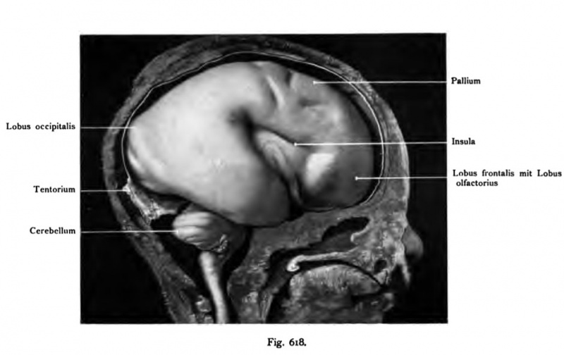
Fig. 618. Brain of a human fetus of 6 months
Fronto-occipital head length ?? cm. Enlarged.
(Anatomical Collection in Basel.)
The brain is shown in situ, DA6 the location of each Ab- sections are marked with the skull and the skull base. the dura mater and the tentorium cerebelli are visible. The Hemisphreres shows the first hint of furrows perpendicular to the center of the island, GE directed to the central sulcus, precentral sulcus and then the slightly right and below the beginning of the superior frontal sulcus. In the lower section the fossa lateralis cerebri (Sylvius) is the embryonic olfactory gyrus lateralis and bent his course to the visible tip of the temporal lobe.
- This text is a Google translate computer generated translation and may contain many errors.
Images from - Atlas of the Development of Man (Volume 2)
(Handatlas der entwicklungsgeschichte des menschen)
- Kollmann Atlas 2: Gastrointestinal | Respiratory | Urogenital | Cardiovascular | Neural | Integumentary | Smell | Vision | Hearing | Kollmann Atlas 1 | Kollmann Atlas 2 | Julius Kollmann
- Links: Julius Kollman | Atlas Vol.1 | Atlas Vol.2 | Embryology History
| Historic Disclaimer - information about historic embryology pages |
|---|
| Pages where the terms "Historic" (textbooks, papers, people, recommendations) appear on this site, and sections within pages where this disclaimer appears, indicate that the content and scientific understanding are specific to the time of publication. This means that while some scientific descriptions are still accurate, the terminology and interpretation of the developmental mechanisms reflect the understanding at the time of original publication and those of the preceding periods, these terms, interpretations and recommendations may not reflect our current scientific understanding. (More? Embryology History | Historic Embryology Papers) |
Reference
Kollmann JKE. Atlas of the Development of Man (Handatlas der entwicklungsgeschichte des menschen). (1907) Vol.1 and Vol. 2. Jena, Gustav Fischer. (1898).
Cite this page: Hill, M.A. (2024, April 19) Embryology Kollmann618.jpg. Retrieved from https://embryology.med.unsw.edu.au/embryology/index.php/File:Kollmann618.jpg
- © Dr Mark Hill 2024, UNSW Embryology ISBN: 978 0 7334 2609 4 - UNSW CRICOS Provider Code No. 00098G
Fig. 618. Gehirn eines menschlichen Fetus vom 6« Monat
Fronto-occipitale Kopflänge ^ cm. Vergrößert. (Anatomische Sammlung in Basel.)
Das Gehirn ist in situ dargestellt, so da6 die Lage der einzelnen Ab- schnitte zu dem Schädeldach und der Schädelbasis kenntlich werden. Die Dura mater encephali und das Tentorium cerebelli sind sichtbar. Das Hemisphärium zeigt die erste Andeutung von Furchen: senkrecht auf die Mitte der Insel ge- richtet den Sulcus centralis, dann den Sulcus praecentralis und etwas nach rechts und unten der Beginn des Sulcus frontalis superior. Im unteren Abschnitt der Fossa cerebri lateralis (Sylvii) ist der embryonale Gyrus olfactorius lateralis und sein gebogener Verlauf zur Spitze des Schläfenlappens sichtbar.
File history
Click on a date/time to view the file as it appeared at that time.
| Date/Time | Thumbnail | Dimensions | User | Comment | |
|---|---|---|---|---|---|
| current | 17:05, 17 October 2011 |  | 940 × 592 (50 KB) | S8600021 (talk | contribs) | {{Kollmann1907}} Category:Human Category:Neural Fig. 618. Gehirn eines menschlichen Fetus vom 6« Monat Fronto-occipitale Kopflänge ^ cm. Vergrößert. (Anatomische Sammlung in Basel.) Das Gehirn ist in situ dargestellt, so da6 die Lag |
You cannot overwrite this file.
File usage
The following page uses this file:
