File:Kollmann614.jpg
Kollmann614.jpg (679 × 502 pixels, file size: 33 KB, MIME type: image/jpeg)
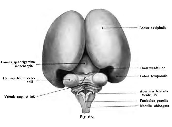
Fig. 614. Human fetus of 4 Month (10 cm CRL)
Seen from behind. The same brain as in Fig. 613.
(Anatomical Collection in Basel.)
The lateral hemisphere areas are still vöUig smooth. A recovery back to the medial surface as well Thalamusmulde caused by adaptation of the hemisphere wall at the convexity of the thalamus. The occipital lobes diverge, making the midbrain (mesencephalon) namely the quadrigeminal plate in large part is visible. The view from behind the cerebellum lies before the observer. His hemispheres stand out clearly as the dorsal surface of the diamond mine lying in front of the gracile funiculi. The cerebrum shows nowhere transient furrows. The fetus was immediately after the expulsion with formol 10: 100 injects, opened after a few days away, the skull and the brain.
- This text is a Google translate computer generated translation and may contain many errors.
Images from - Atlas of the Development of Man (Volume 2)
(Handatlas der entwicklungsgeschichte des menschen)
- Kollmann Atlas 2: Gastrointestinal | Respiratory | Urogenital | Cardiovascular | Neural | Integumentary | Smell | Vision | Hearing | Kollmann Atlas 1 | Kollmann Atlas 2 | Julius Kollmann
- Links: Julius Kollman | Atlas Vol.1 | Atlas Vol.2 | Embryology History
| Historic Disclaimer - information about historic embryology pages |
|---|
| Pages where the terms "Historic" (textbooks, papers, people, recommendations) appear on this site, and sections within pages where this disclaimer appears, indicate that the content and scientific understanding are specific to the time of publication. This means that while some scientific descriptions are still accurate, the terminology and interpretation of the developmental mechanisms reflect the understanding at the time of original publication and those of the preceding periods, these terms, interpretations and recommendations may not reflect our current scientific understanding. (More? Embryology History | Historic Embryology Papers) |
Reference
Kollmann JKE. Atlas of the Development of Man (Handatlas der entwicklungsgeschichte des menschen). (1907) Vol.1 and Vol. 2. Jena, Gustav Fischer. (1898).
Cite this page: Hill, M.A. (2024, April 24) Embryology Kollmann614.jpg. Retrieved from https://embryology.med.unsw.edu.au/embryology/index.php/File:Kollmann614.jpg
- © Dr Mark Hill 2024, UNSW Embryology ISBN: 978 0 7334 2609 4 - UNSW CRICOS Provider Code No. 00098G
Fig. 614. Qehirn eines menschlichen Fetus vom 4. Monat (10 cm Scheitel-
steifilänse).
Von hinten gesehen. Dasselbe Gehirn wie in Fig. 613.
(Anatomische Sammlung in Basel.)
Die lateralen Hemisphärenflächen sind noch vöUig glatt. Eine Einziehung hinten an der medialen Fläche ist als Thalamusmulde wohl durch Anpassung der Hemisphärenwand an die Konvexität des Thalamus entstanden. Die Lobi occipitales weichen auseinander, wodurch das Mittelhirn (Mesencephalon) und zwar dessen Vierhügelplatte zu einem großen Teile sichtbar ist.
Bei der Ansicht von hinten liegt das Kleinhirn vor dem Beschauer. Seine Hemisphären treten deutlich hervor, ebenso die dorsale Fläche der Rauten- grube vor den Funiculi graciles liegend.
Das Großhirn zeigt nirgends transitorische Furchen. Der Fetus wurde direkt nach der Ausstoßung mit Formol 10 : 100 injiziert, nach ein paar Tagen der Schädel geöffnet und das Gehirn entfernt.
File history
Click on a date/time to view the file as it appeared at that time.
| Date/Time | Thumbnail | Dimensions | User | Comment | |
|---|---|---|---|---|---|
| current | 17:02, 17 October 2011 |  | 679 × 502 (33 KB) | S8600021 (talk | contribs) | {{Kollmann1907}} Category:Human Category:Neural Fig. 614. Qehirn eines menschlichen Fetus vom 4. Monat (10 cm Scheitel- steifilänse). Von hinten gesehen. Dasselbe Gehirn wie in Fig. 613. (Anatomische Sammlung in Basel.) Die lateralen |
You cannot overwrite this file.
File usage
The following page uses this file:

