File:Kollmann613.jpg
Kollmann613.jpg (652 × 506 pixels, file size: 31 KB, MIME type: image/jpeg)
- This text is a Google translate computer generated translation and may contain many errors.
Images from - Atlas of the Development of Man (Volume 2)
(Handatlas der entwicklungsgeschichte des menschen)
- Kollmann Atlas 2: Gastrointestinal | Respiratory | Urogenital | Cardiovascular | Neural | Integumentary | Smell | Vision | Hearing | Kollmann Atlas 1 | Kollmann Atlas 2 | Julius Kollmann
- Links: Julius Kollman | Atlas Vol.1 | Atlas Vol.2 | Embryology History
| Historic Disclaimer - information about historic embryology pages |
|---|
 |
Reference
Kollmann JKE. Atlas of the Development of Man (Handatlas der entwicklungsgeschichte des menschen). (1907) Vol.1 and Vol. 2. Jena, Gustav Fischer. (1898).
Cite this page: Hill, M.A. (2024, April 24) Embryology Kollmann613.jpg. Retrieved from https://embryology.med.unsw.edu.au/embryology/index.php/File:Kollmann613.jpg
- © Dr Mark Hill 2024, UNSW Embryology ISBN: 978 0 7334 2609 4 - UNSW CRICOS Provider Code No. 00098G
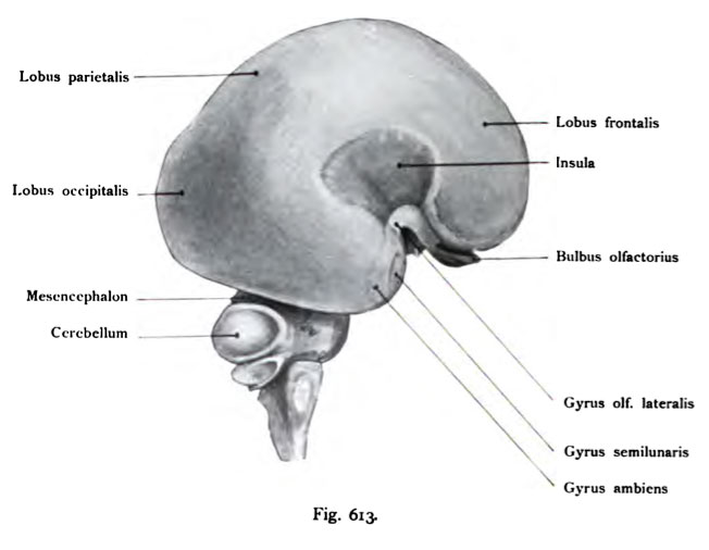
Fig. 613. Qehirn eines menschlichen Fetus vom Anfang des 4. Monates,
von der lateralen Fläche gesehen. Vergr. 4 mal. (Anatomische Samndung in Basel)
Die Oberfläche des Pallium ist auf der linken Seite noch glatt, die früher beobachteten transitorischen Furchen sind als postmortale Erscheinungen erkannt worden. Das Hemisphärium wiederholt den Zustand der lissencephalen Ge- hirne der Säuger, Es hat gegen den 3. Monat (Fig. 608) bedeutend an Umfang zugenommen und bedeckt mit den Lobi occipitales das Mittelhirn fast vollständig. Die Fossa cerebri lateralis (Sylvii) ist als ein vertieftes birnförmiges Feld deut- lich ausgeprägt. Oral zieht der Gyrus lateralis rhinencephali zur Spitze des Schläfenlappens in Form eines stark gebogenen Streifens ; die Stelle der stärksten Krümmung heißt Angulus gyri olfactorii lateralis. Dieser laterale Gyrus olfac- torius tritt in den G3rrus ambiens und Gyrus semilunaris ein. Das Kleinhirn ist noch wenig entwickelt.
File history
Click on a date/time to view the file as it appeared at that time.
| Date/Time | Thumbnail | Dimensions | User | Comment | |
|---|---|---|---|---|---|
| current | 17:01, 17 October 2011 | | 652 × 506 (31 KB) | S8600021 (talk | contribs) | {{Kollmann1907}} Category:Human Category:Neural Fig. 613. Qehirn eines menschlichen Fetus vom Anfang des 4. Monates, von der lateralen Fläche gesehen. Vergr. 4 mal. (Anatomische Samndung in Basel) Die Oberfläche des Pallium ist auf der |
You cannot overwrite this file.
File usage
The following page uses this file: