File:Kollmann608.jpg
Kollmann608.jpg (697 × 563 pixels, file size: 77 KB, MIME type: image/jpeg)
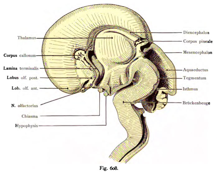
Fig. 608. Brain of a fetus in three monthly median section
(Anatomical Collection in Basel)
The median surface of the right hemisphere exposed. Inside, the development has increased in each institution. Ventriculus tertius in the thalamus and hypothalamus are visible. The spread of the pallium, the rhinencephalon is sometimes pushed to the base and the medial surface of the Hemisphärium show how the olfactory lobe anterior and posterior. Apart from the above mentioned sections, the N. olfactorius, the pineal lamina terminalis, the corpus callosum, corpus striatum and inter-ventricular foramen (of Monro), the roof of the diencephalon, the investment of the corpus of the mesencephalon, aqueduct, and the Isthmus bridge curvature highlighted by labels. The corpus callosum 2 V «mm in size, is still ahead of the thalamus. Splenium, corpus, Genu, Rostrum are recognizable despite the smallness.
- This text is a Google translate computer generated translation and may contain many errors.
Images from - Atlas of the Development of Man (Volume 2)
(Handatlas der entwicklungsgeschichte des menschen)
- Kollmann Atlas 2: Gastrointestinal | Respiratory | Urogenital | Cardiovascular | Neural | Integumentary | Smell | Vision | Hearing | Kollmann Atlas 1 | Kollmann Atlas 2 | Julius Kollmann
- Links: Julius Kollman | Atlas Vol.1 | Atlas Vol.2 | Embryology History
| Historic Disclaimer - information about historic embryology pages |
|---|
| Pages where the terms "Historic" (textbooks, papers, people, recommendations) appear on this site, and sections within pages where this disclaimer appears, indicate that the content and scientific understanding are specific to the time of publication. This means that while some scientific descriptions are still accurate, the terminology and interpretation of the developmental mechanisms reflect the understanding at the time of original publication and those of the preceding periods, these terms, interpretations and recommendations may not reflect our current scientific understanding. (More? Embryology History | Historic Embryology Papers) |
Reference
Kollmann JKE. Atlas of the Development of Man (Handatlas der entwicklungsgeschichte des menschen). (1907) Vol.1 and Vol. 2. Jena, Gustav Fischer. (1898).
Cite this page: Hill, M.A. (2024, April 25) Embryology Kollmann608.jpg. Retrieved from https://embryology.med.unsw.edu.au/embryology/index.php/File:Kollmann608.jpg
- © Dr Mark Hill 2024, UNSW Embryology ISBN: 978 0 7334 2609 4 - UNSW CRICOS Provider Code No. 00098G
Fig. 608. Gehirn eines 3 monatlichen Fetus im Medianschnitt.
(Anatomische Sammlung in Basel)
Die mediane Fläche des rechten Hemisphärium liegt frei. Im Innern hat die Entwicklung der einzelnen Organe zugenommen. In dem Ventriculus tertius sind Thalamus und Hypothalamus sichtbar. Durch die Ausbreitung des Pallium ist das Rhinencephalon teilweise an die Basis und die mediale Fläche des Hemisphärium gedrängt, wie der Lobus olfactorius anterior und posterior zeigen. Abgesehen von den ebenerwähnten Abschnitten ist der N. olfactorius, die Lamina terminalis, das Corpus callosum, Corpus striatum und Foramen inter- ventriculare (Monroi), das Dach des Diencephalon , die Anlage des Corpus pineale, des Mesencephalon, Aquaeductus, Isthmus und die Brückenkrümmung durch Bezeichnungen hervorgehoben. Das Corpus callosum ist 2 V« mm groß, liegt noch vor dem Thalamus. Splenium, Corpus, Genu, Rostrum sind trotz der Kleinheit erkennbar.
File history
Click on a date/time to view the file as it appeared at that time.
| Date/Time | Thumbnail | Dimensions | User | Comment | |
|---|---|---|---|---|---|
| current | 16:56, 17 October 2011 |  | 697 × 563 (77 KB) | S8600021 (talk | contribs) | {{Kollmann1907}} Category:Human Category:Neural |
You cannot overwrite this file.
File usage
The following page uses this file:

