File:Kollmann534.jpg
Kollmann534.jpg (684 × 599 pixels, file size: 74 KB, MIME type: image/jpeg)
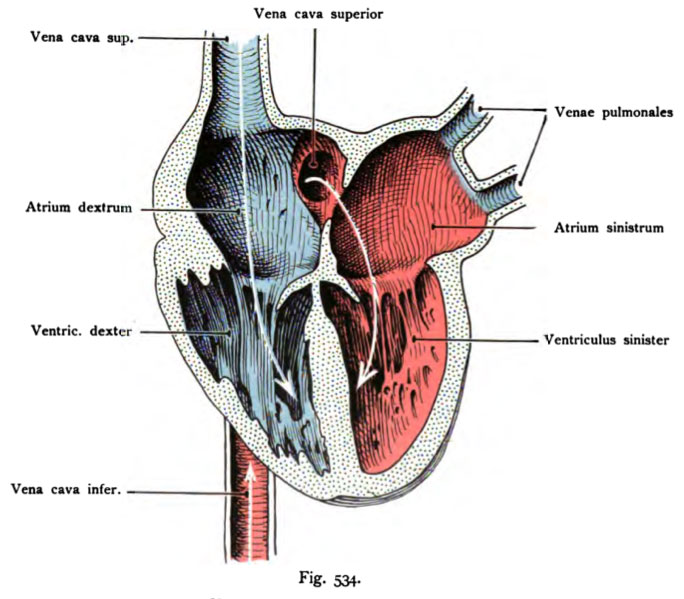
Fig. 534. Fetal heart, dorsal half with the afferent paths, open and colored according to the physiological condition of the blood
semi-schematically and enlarged.
(After Boom.)
The heart is shown in diastole. The vena cava. brings venous blood that pours through the atrium into the right ventricle. The inferior vena cava brings blood mixed, so painted red. The blood passes from the inferior vena cava through the foramen ovale into the left atrium, then left the chamber. The left atrium receives venous blood still through the pulmonary veins. White arrows indicate the flow direction.
- This text is a Google translate computer generated translation and may contain many errors.
Images from - Atlas of the Development of Man (Volume 2)
(Handatlas der entwicklungsgeschichte des menschen)
- Kollmann Atlas 2: Gastrointestinal | Respiratory | Urogenital | Cardiovascular | Neural | Integumentary | Smell | Vision | Hearing | Kollmann Atlas 1 | Kollmann Atlas 2 | Julius Kollmann
- Links: Julius Kollman | Atlas Vol.1 | Atlas Vol.2 | Embryology History
| Historic Disclaimer - information about historic embryology pages |
|---|
| Pages where the terms "Historic" (textbooks, papers, people, recommendations) appear on this site, and sections within pages where this disclaimer appears, indicate that the content and scientific understanding are specific to the time of publication. This means that while some scientific descriptions are still accurate, the terminology and interpretation of the developmental mechanisms reflect the understanding at the time of original publication and those of the preceding periods, these terms, interpretations and recommendations may not reflect our current scientific understanding. (More? Embryology History | Historic Embryology Papers) |
Reference
Kollmann JKE. Atlas of the Development of Man (Handatlas der entwicklungsgeschichte des menschen). (1907) Vol.1 and Vol. 2. Jena, Gustav Fischer. (1898).
Cite this page: Hill, M.A. (2024, April 24) Embryology Kollmann534.jpg. Retrieved from https://embryology.med.unsw.edu.au/embryology/index.php/File:Kollmann534.jpg
- © Dr Mark Hill 2024, UNSW Embryology ISBN: 978 0 7334 2609 4 - UNSW CRICOS Provider Code No. 00098G
Fig. 534. Fetales Herz, dorsale Hälfte mit den zuführenden Bahnen , geöffnet
und entsprechend der physiologischen Beschaffenheit des Blutes koloriert. Halb- schematisch und vergrößert.
(Nach Bumm.)
Das Herz ist in der Diastole dargestellt. Die Vena cava sup. bringt venöses Blut, das sich durch den Vorhof in die rechte Kammer ergießt. Die Vena cava inferior bringt gemischtes Blut, deshalb rot bemalt. Das Blut gelangt von der Vena cava inferior durch das Foramen ovale in den linken Vor- hof, dann in die linke Kammer. Der linke Vorhof erhält noch venöses Blut durch die Lungenvenen. Weiße Pfeile deuten auf die Stromrichtung.
File history
Click on a date/time to view the file as it appeared at that time.
| Date/Time | Thumbnail | Dimensions | User | Comment | |
|---|---|---|---|---|---|
| current | 00:09, 17 October 2011 |  | 684 × 599 (74 KB) | S8600021 (talk | contribs) | {{Kollmann1907}} |
You cannot overwrite this file.
File usage
The following 2 pages use this file:

