File:Kollmann532.jpg
Kollmann532.jpg (702 × 409 pixels, file size: 59 KB, MIME type: image/jpeg)
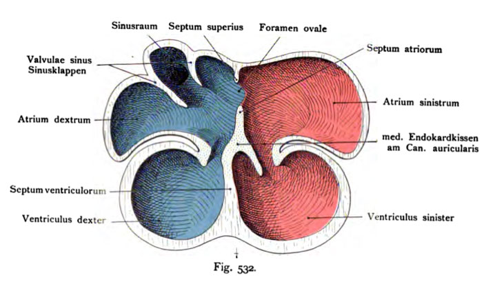
Fig. 532. Development of the heart chambers and septa
Last stage. View the dorsal half. Schematically. (According to Hochstetter. From the manual of the history of O. Hartwig.)
The papillary muscles and trabeculae are carneae omitted. the Interventricular septum atriorum is now the heart of vaginal septum wall (atrio-ventricular septum), united with the medial endocardial cushion the former Canalis auricularis convey the connection. You and the septum atriorum are dotted. The medial endocardial cushion also supply some of material for the production of the atrioventricular valves. In the septum atrium, at its root, a single larger upper opening-up occurs, the foramen ovale. The septum is superior later its sickle-shaped closure.
- This text is a Google translate computer generated translation and may contain many errors.
Images from - Atlas of the Development of Man (Volume 2)
(Handatlas der entwicklungsgeschichte des menschen)
- Kollmann Atlas 2: Gastrointestinal | Respiratory | Urogenital | Cardiovascular | Neural | Integumentary | Smell | Vision | Hearing | Kollmann Atlas 1 | Kollmann Atlas 2 | Julius Kollmann
- Links: Julius Kollman | Atlas Vol.1 | Atlas Vol.2 | Embryology History
| Historic Disclaimer - information about historic embryology pages |
|---|
| Pages where the terms "Historic" (textbooks, papers, people, recommendations) appear on this site, and sections within pages where this disclaimer appears, indicate that the content and scientific understanding are specific to the time of publication. This means that while some scientific descriptions are still accurate, the terminology and interpretation of the developmental mechanisms reflect the understanding at the time of original publication and those of the preceding periods, these terms, interpretations and recommendations may not reflect our current scientific understanding. (More? Embryology History | Historic Embryology Papers) |
Reference
Kollmann JKE. Atlas of the Development of Man (Handatlas der entwicklungsgeschichte des menschen). (1907) Vol.1 and Vol. 2. Jena, Gustav Fischer. (1898).
Cite this page: Hill, M.A. (2024, April 24) Embryology Kollmann532.jpg. Retrieved from https://embryology.med.unsw.edu.au/embryology/index.php/File:Kollmann532.jpg
- © Dr Mark Hill 2024, UNSW Embryology ISBN: 978 0 7334 2609 4 - UNSW CRICOS Provider Code No. 00098G
Fig. 532. Ausgestaltung des Herzinnern. Entwicklung der Scheidewände.
Letzte Stufe. Ansicht der dorsalen Hälfte. Schematisch. (Nach Hochstetter. Aus dem Handbuch der Entwicklungsgeschichte von O. Hartwig.)
Die Papillär muskeln und die Trabeculae carneae sind weggelassen. Das Septum atriorum ist jetzt mit dem Septum interventriculare zur Herzscheiden- wand (Septum atrio-ventriculare) vereinigt, wobei die medialen Endokardkissen des früheren Canalis auricularis die Verbindung vermitteln. Sie und das Septum atriorum sind punktiert. Die medialen Endokardkissen liefern auch zum Teil Material für die Herstellung der Atrioventrikularklappen. In dem Septum atriorum ist an seiner oberen Wurzel eine einheitliche größere Öffnung aufge- treten, das Foramen ovale. Das Septum superius bildet später dessen sichel- förmigen Abschluß.
File history
Click on a date/time to view the file as it appeared at that time.
| Date/Time | Thumbnail | Dimensions | User | Comment | |
|---|---|---|---|---|---|
| current | 00:09, 17 October 2011 |  | 702 × 409 (59 KB) | S8600021 (talk | contribs) | {{Kollmann1907}} |
You cannot overwrite this file.
File usage
The following 2 pages use this file:

