File:Kollmann529.jpg
Kollmann529.jpg (737 × 593 pixels, file size: 94 KB, MIME type: image/jpeg)
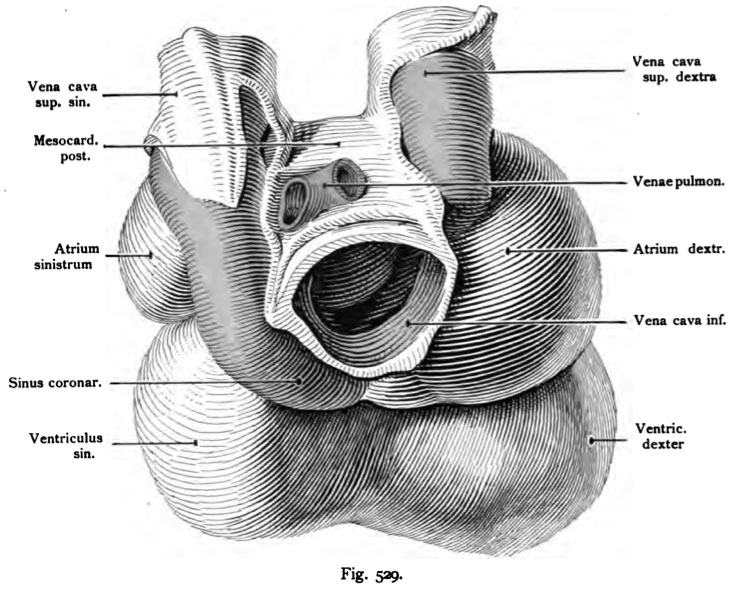
Fig. 529. The heart of a 24 mm Embryo
Seen from behind.
A portion of the vein, the former from the rear into the two atria, one-penetrated, Fig 527, have received other compounds, such as the vena umbilicalis artery, which is converted into a vein of the abdominal wall, or how the
Omphalomesenterica veins, which, like the umbilical vein into the vena Now enter the liver, etc. are only directly in connection with the heart: The two superior vena cava, the inferior vena cava and the veins pulmonary, which enter through the posterior mesocardium.
- This text is a Google translate computer generated translation and may contain many errors.
Images from - Atlas of the Development of Man (Volume 2)
(Handatlas der entwicklungsgeschichte des menschen)
- Kollmann Atlas 2: Gastrointestinal | Respiratory | Urogenital | Cardiovascular | Neural | Integumentary | Smell | Vision | Hearing | Kollmann Atlas 1 | Kollmann Atlas 2 | Julius Kollmann
- Links: Julius Kollman | Atlas Vol.1 | Atlas Vol.2 | Embryology History
| Historic Disclaimer - information about historic embryology pages |
|---|
 |
Reference
Kollmann JKE. Atlas of the Development of Man (Handatlas der entwicklungsgeschichte des menschen). (1907) Vol.1 and Vol. 2. Jena, Gustav Fischer. (1898).
Cite this page: Hill, M.A. (2024, April 18) Embryology Kollmann529.jpg. Retrieved from https://embryology.med.unsw.edu.au/embryology/index.php/File:Kollmann529.jpg
- © Dr Mark Hill 2024, UNSW Embryology ISBN: 978 0 7334 2609 4 - UNSW CRICOS Provider Code No. 00098G
Fig. 529. Das Herz eines Meosclieoeinbryo von 24 mm Sclieitelsteifilänge
von hinten gesehen.
Ein Teil der Venen, die früher von hinten her in die beiden Atrien ein- drangen, Fig. 527, haben andere Verbindungen erhalten, wie die Vena umbili- calis dextra, die zu einer Vene der Bauch wand umgewandelt ist, oder wie die Venae omphalo-mesentericae, welche wie die Vena umbilicalis sinistra in die Leber eintreten usw. Jetzt sind nur noch direkt mit dem Herzen in Verbindung: Die beiden Venae cavae superiores, die Vena cava inferior und die Venae pul- monales, welche durch das Mesocardium posterius eintreten.
File history
Click on a date/time to view the file as it appeared at that time.
| Date/Time | Thumbnail | Dimensions | User | Comment | |
|---|---|---|---|---|---|
| current | 23:34, 16 October 2011 | | 737 × 593 (94 KB) | S8600021 (talk | contribs) | {{Kollmann1907}} |
You cannot overwrite this file.
File usage
The following 2 pages use this file: