File:Kollmann528.jpg
Kollmann528.jpg (744 × 587 pixels, file size: 91 KB, MIME type: image/jpeg)
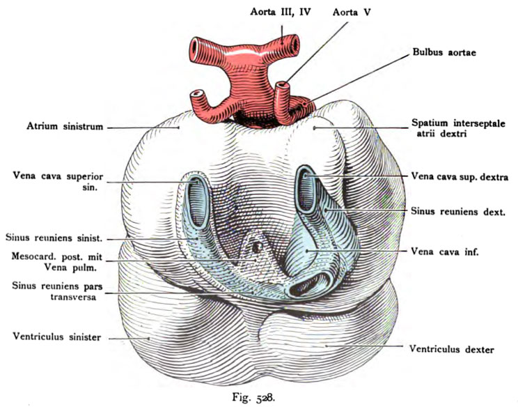
Fig. 528. Heart of a Rabbit Embryo seen from behind at 3.4 mm head length
(from Vorderhim and that of the most prominent point up. for above item the Mittelhims measured). 12V2 days after mating.
(According to one model of Born.)
At the posterior part of the embryonic heart come the large veins one, are shown here:
The two superior vena cava (artery and vein).
The vena cava inferior and the sinus reuniens dexter and sinister with the across the bottom floor, causing the left pours his blood into the right. the dotted surface on the wall of the sinus and between the same shows the coalescence of the posterior mesocardium with the system and lung to the pulmonary vein. The antechamber of the heart by a deep, broad bay externally separated into right and left half. with the Relocation of the atria are the ductus and the vena cava Cuvieri. moved into the air (see Fig 525).
- This text is a Google translate computer generated translation and may contain many errors.
Images from - Atlas of the Development of Man (Volume 2)
(Handatlas der entwicklungsgeschichte des menschen)
- Kollmann Atlas 2: Gastrointestinal | Respiratory | Urogenital | Cardiovascular | Neural | Integumentary | Smell | Vision | Hearing | Kollmann Atlas 1 | Kollmann Atlas 2 | Julius Kollmann
- Links: Julius Kollman | Atlas Vol.1 | Atlas Vol.2 | Embryology History
| Historic Disclaimer - information about historic embryology pages |
|---|
| Pages where the terms "Historic" (textbooks, papers, people, recommendations) appear on this site, and sections within pages where this disclaimer appears, indicate that the content and scientific understanding are specific to the time of publication. This means that while some scientific descriptions are still accurate, the terminology and interpretation of the developmental mechanisms reflect the understanding at the time of original publication and those of the preceding periods, these terms, interpretations and recommendations may not reflect our current scientific understanding. (More? Embryology History | Historic Embryology Papers) |
Reference
Kollmann JKE. Atlas of the Development of Man (Handatlas der entwicklungsgeschichte des menschen). (1907) Vol.1 and Vol. 2. Jena, Gustav Fischer. (1898).
Cite this page: Hill, M.A. (2024, April 24) Embryology Kollmann528.jpg. Retrieved from https://embryology.med.unsw.edu.au/embryology/index.php/File:Kollmann528.jpg
- © Dr Mark Hill 2024, UNSW Embryology ISBN: 978 0 7334 2609 4 - UNSW CRICOS Provider Code No. 00098G
Fig. 528. Herz eines Kanhichenembryo von hinten gesehen bei 3,4 mm Kopflänge
(vom Vorderhim und zwar vom vorstehendsten Punkt bis. zum vorstehenden Punkt des Mittelhims gemessen). 12V2 Tage nach der Begattung.
(Nach einem Modell von Born.)
Am hinteren Umfang des embryonalen Herzens treten die großen Venen ein; hier sind dargestellt:
Die beiden Venae cavae superiores (dextra und sinistra).
Die Vena cava inferior und der Sinus reuniens dexter und sinister mit dem unteren QuerstOck, wodurch der linke sein Blut in den rechten ergießt. Die punktierte Oberfläche an der Wand der Sinus und zwischen denselben zeigt die Verwachsung durch das Mesocardium posterius mit der Lungenanlage und die Lungenvene dazu. Die Vorkammerabteilung des Herzens wird durch eine tiefe, breite Bucht äußerlich in eine rechte und linke Hälfte getrennt. Mit der Verlagerung der Vorhöfe sind auch die Ductus Cuvieri und die Vena cava inf. in die Höhe gerückt (vergl. Fig. 525).
File history
Click on a date/time to view the file as it appeared at that time.
| Date/Time | Thumbnail | Dimensions | User | Comment | |
|---|---|---|---|---|---|
| current | 23:34, 16 October 2011 |  | 744 × 587 (91 KB) | S8600021 (talk | contribs) | {{Kollmann1907}} |
You cannot overwrite this file.
File usage
The following 2 pages use this file:

