File:Kollmann512.jpg
Original file (737 × 842 pixels, file size: 100 KB, MIME type: image/jpeg)
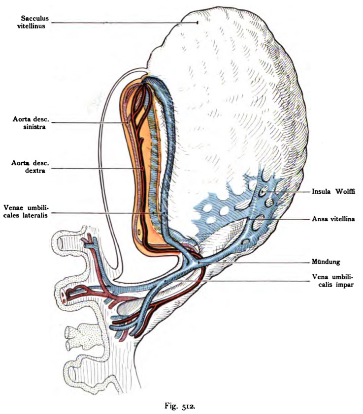
Fig. 512. Vascular system of a human embryo of 1.3 mm in length
Schematic lateral view of the embryo shown in Fig. 511.
(After Eternod.)
The heart is located directly in front of the germinal disc in the boundary of the Yolk sac. From the heart arise from two aortas. Each aorta has two arterial arch, and a third is still rudimentary. The two aortic run separately on each side of the notochord to the rear, without connect and go through the belly stalk separated by the chorionic villi.
The chorionic villi from the umbilical veins returning Primitiva unite in the abdominal peduncle a short trunk, inferior vena umbilicalis impar, which follows the Allantoisgang. Shares after a short course in the unpaired base in two large veins, which run close to the marginal ridge of the yolk sac to the front of the yellow pages of the embryo Alfeld. These two lateral umbilical veins split in the rear Heart thighs, this horseshoe-shaped grip around the head end of the Area embryonalis. The umbilical veins lateral caudal to take two vitelline veins veins vitellina on. They arise from the wide capillaries that the yolk sac islands (Wolff) surrounded. These veins vitellina Ansa vitellina form a loop around the mouth of the allantois.
- This text is a Google translate computer generated translation and may contain many errors.
Images from - Atlas of the Development of Man (Volume 2)
(Handatlas der entwicklungsgeschichte des menschen)
- Kollmann Atlas 2: Gastrointestinal | Respiratory | Urogenital | Cardiovascular | Neural | Integumentary | Smell | Vision | Hearing | Kollmann Atlas 1 | Kollmann Atlas 2 | Julius Kollmann
- Links: Julius Kollman | Atlas Vol.1 | Atlas Vol.2 | Embryology History
| Historic Disclaimer - information about historic embryology pages |
|---|
 |
Reference
Kollmann JKE. Atlas of the Development of Man (Handatlas der entwicklungsgeschichte des menschen). (1907) Vol.1 and Vol. 2. Jena, Gustav Fischer. (1898).
Cite this page: Hill, M.A. (2024, April 23) Embryology Kollmann512.jpg. Retrieved from https://embryology.med.unsw.edu.au/embryology/index.php/File:Kollmann512.jpg
- © Dr Mark Hill 2024, UNSW Embryology ISBN: 978 0 7334 2609 4 - UNSW CRICOS Provider Code No. 00098G
Fig. 512. Qefäßsystem eines menschlichen Embryo von 1,3 mm Länge.
Schema. Norma lateralis des Embryo Fig. 511.
(Nach E tarn od.)
Das Herz befindet sich unmittelbar vor der Keimscheibe in dem Rand des Dottersackes. Aus dem Herz entspringen zwei Aorten. Jede Aorta besitzt zwei arterielle Bogen, ein dritter ist noch rudimentär. Die beiden Aorten verlaufen zu beiden Seiten der Chordaanlage getrennt nach hinten, ohne sich zu verbinden und gehen getrennt durch den Bauchstiel nach den Chorionzotten.
Die aus den Chorionzotten zurückkehrenden Venae umbilicales primitivae vereinigen sich im Pedunculus abdominalis zu einem kurzen Stamm, Vena umbilicalis impar, der dem Allantoisgang folgt. Nach kurzem Verlauf teilt sich der unpaare Stamm in zwei große Venen, welche in dem Randwulst des Dottersackes nach vorn ziehen dicht an den Seiten des gelben Embryonalfeldes. Diese beiden Venae umbilicales laterales ergießen sich getrennt in die hinteren Herzschenkel, diese umgreife^ hufeisenförmig das Kopfende der Area embryonalis. Die Venae umbilicales laterales nehmen kaudal zwei Dottervenen Venae vitellinae auf. Sie entstehen aus den weiten Kapillaren, welche die Dottersackinseln (Wolff) umgeben. Diese Venae vitellinae bilden eine Schlinge Ansa vitellina um die Mündung des AUantoisganges.
File history
Click on a date/time to view the file as it appeared at that time.
| Date/Time | Thumbnail | Dimensions | User | Comment | |
|---|---|---|---|---|---|
| current | 23:16, 16 October 2011 | | 737 × 842 (100 KB) | S8600021 (talk | contribs) | {{Kollmann1907}} |
You cannot overwrite this file.
File usage
The following 2 pages use this file: