File:Kollmann511.jpg

Original file (710 × 838 pixels, file size: 108 KB, MIME type: image/jpeg)
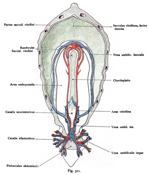
Fig. 511. Vascular system of a human embryo of 1.3 mm
Schematic ventral view.
(After Eternod.)
The yolk sac is removed at the point of highest circumference.
One sees in its interior. The thickness of the yolk sac wall are blood vessels indicated, as are the layer of the endoderm and in the middle
Orientation of the chordal and embryonalis Area.
The arterial system consists of two and two aortic arches, Aortenende spring from the heart. A third form is the develop- are understood. The aorta descendents run separately developed the notochord long stem and through the abdomen in order to spread throughout the chorionic villi. the returning veins are veins chorio-placental, two of them go Umbilical veins produce. These unite in the abdominal peduncle to an impar umbilical vein, which after a short course again in two Lateral umbilical vein separates, which side of the area in embryonalis the marginal ridge of the yolk sac included the horseshoe-shaped Femoral veins of the heart draw. The lateral umbilical veins take near the peduncle, the Ansa abdominahs vitellina (the two venae vitellina from the circle to venosus).
- This text is a Google translate computer generated translation and may contain many errors.
Images from - Atlas of the Development of Man (Volume 2)
(Handatlas der entwicklungsgeschichte des menschen)
- Kollmann Atlas 2: Gastrointestinal | Respiratory | Urogenital | Cardiovascular | Neural | Integumentary | Smell | Vision | Hearing | Kollmann Atlas 1 | Kollmann Atlas 2 | Julius Kollmann
- Links: Julius Kollman | Atlas Vol.1 | Atlas Vol.2 | Embryology History
| Historic Disclaimer - information about historic embryology pages |
|---|
| Pages where the terms "Historic" (textbooks, papers, people, recommendations) appear on this site, and sections within pages where this disclaimer appears, indicate that the content and scientific understanding are specific to the time of publication. This means that while some scientific descriptions are still accurate, the terminology and interpretation of the developmental mechanisms reflect the understanding at the time of original publication and those of the preceding periods, these terms, interpretations and recommendations may not reflect our current scientific understanding. (More? Embryology History | Historic Embryology Papers) |
Reference
Kollmann JKE. Atlas of the Development of Man (Handatlas der entwicklungsgeschichte des menschen). (1907) Vol.1 and Vol. 2. Jena, Gustav Fischer. (1898).
Cite this page: Hill, M.A. (2024, April 24) Embryology Kollmann511.jpg. Retrieved from https://embryology.med.unsw.edu.au/embryology/index.php/File:Kollmann511.jpg
- © Dr Mark Hill 2024, UNSW Embryology ISBN: 978 0 7334 2609 4 - UNSW CRICOS Provider Code No. 00098G
Fig. 511. Qefäßsystem eines menschlichen Embryo von 1,3 mm.
Schema. Norma ventralis. (Nach Eternod.)
Der Dottersack ist auf der Stelle der höchsten Zirkumferenz abgetragen. Man sieht in dessen Innenraum. In der Dicke der Dottersack wand sind Blut- gefäße angegeben, ebenso die Schichte des Entoderms und in der Mitte zur Orientierung die Chordaplatte und die Area embryonalis.
Das arterielle System besteht aus zwei Aorten und zwei Bogen, welche aus dem Aortenende des Herzens entspringen. Ein dritter Bogen ist im Ent- stehen begriffen. Die Aortae descendentes verlaufen getrennt der Chorda ent- lang und durch den Bauchstiel, um sich in den Chorionzotten zu verbreiten. Die zurückkehrenden Venen sind Venae chorio-placentares; aus ihnen gehen zwei Venae umbilicalis hervor. Diese vereinigen sich im Pedunculus abdominalis zu einer Vena umbilicalis impar, die sich nach kurzem Verlaufe wieder in zwei Venae umbilicales laterales trennt, welche zur Seite der Area embryonalis in der Randwulst des Dottersackes eingeschlossen, zum hufeisenförmig geformten Venenschenkel des Herzens ziehen. Die Venae umbilicales laterales nehmen in der Nähe des Pedunculus abdominahs die Ansa vitellina (die zwei Venae vitellinae aus dem Circulus venosus) auf.
File history
Click on a date/time to view the file as it appeared at that time.
| Date/Time | Thumbnail | Dimensions | User | Comment | |
|---|---|---|---|---|---|
| current | 23:16, 16 October 2011 |  | 710 × 838 (108 KB) | S8600021 (talk | contribs) | {{Kollmann1907}} |
You cannot overwrite this file.
File usage
The following 2 pages use this file:
