File:Kollmann448.jpg
Kollmann448.jpg (643 × 576 pixels, file size: 43 KB, MIME type: image/jpeg)
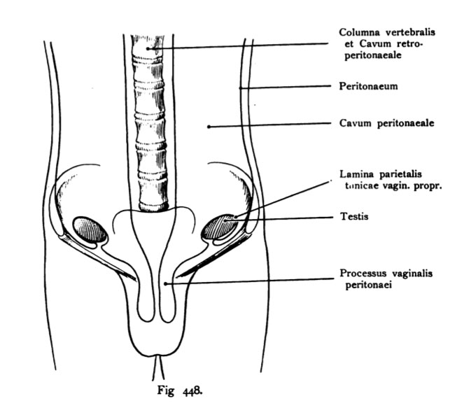
Fig. 448. Testicular descent
Schema. (With changes to Tillaux.)
It shows the emergence of the lamina parietalis of the tunica vaginalis propria at the entrance of the testis into the inguinal canal. The descended testis obtained at the entrance to the inguinal canal, a second coating, the first representing a wide pocket that surrounds him, but later than close and lamina is called the parietal tunica vaginalis propria. The protuberance of the Peritoneum is: processus vaginalis peritonaei. The testicle is in this Figure shows how he gets close to the protuberance of the inguinal canal abdominal inguinal canal to the Apertura and the associated Evagination of the peritoneum is reached. One point of the testis is never from the peritoneum covered, where the blood vessels leak and off.
- This text is a Google translate computer generated translation and may contain many errors.
Images from - Atlas of the Development of Man (Volume 2)
(Handatlas der entwicklungsgeschichte des menschen)
- Kollmann Atlas 2: Gastrointestinal | Respiratory | Urogenital | Cardiovascular | Neural | Integumentary | Smell | Vision | Hearing | Kollmann Atlas 1 | Kollmann Atlas 2 | Julius Kollmann
- Links: Julius Kollman | Atlas Vol.1 | Atlas Vol.2 | Embryology History
| Historic Disclaimer - information about historic embryology pages |
|---|
| Pages where the terms "Historic" (textbooks, papers, people, recommendations) appear on this site, and sections within pages where this disclaimer appears, indicate that the content and scientific understanding are specific to the time of publication. This means that while some scientific descriptions are still accurate, the terminology and interpretation of the developmental mechanisms reflect the understanding at the time of original publication and those of the preceding periods, these terms, interpretations and recommendations may not reflect our current scientific understanding. (More? Embryology History | Historic Embryology Papers) |
Reference
Kollmann JKE. Atlas of the Development of Man (Handatlas der entwicklungsgeschichte des menschen). (1907) Vol.1 and Vol. 2. Jena, Gustav Fischer. (1898).
Cite this page: Hill, M.A. (2024, April 23) Embryology Kollmann448.jpg. Retrieved from https://embryology.med.unsw.edu.au/embryology/index.php/File:Kollmann448.jpg
- © Dr Mark Hill 2024, UNSW Embryology ISBN: 978 0 7334 2609 4 - UNSW CRICOS Provider Code No. 00098G
Fig. 448. Descensus testicuionim.
Schema. (Mit Änderungen nach Tillaux.)
Es zeigt die Entstehung der Lamina parietalis der Tunica vaginalis propria bei dem Eintritt des Hodens in den Leistenkanal. Der herabgestiegene Hoden erhält bei dem Eintritt in den Leistenkanal einen zweiten Überzug, der anfangs eine weite Tasche darstellt, die ihn aber später enge umschließt und als Lamina parietalis der Tunica vaginalis propria bezeichnet wird. Die Ausstülpung des Peritoneums heißt: Processus vaginalis peritonaei. Der Hoden ist in dieser Abbildung dargestellt, wie er in die Nähe der Ausstülpung des Leistenkanales an der Apertura canalis inguinalis abdominalis und an der damit verbundenen Ausstülpung des Peritoneums angelangt ist. Eine Stelle des Hodens wird nie vom Peritonaeum überzogen, dort, wo die Gefäße ein- und austreten.
File history
Click on a date/time to view the file as it appeared at that time.
| Date/Time | Thumbnail | Dimensions | User | Comment | |
|---|---|---|---|---|---|
| current | 21:46, 16 October 2011 |  | 643 × 576 (43 KB) | S8600021 (talk | contribs) | {{Kollmann1907}} |
You cannot overwrite this file.
File usage
The following 3 pages use this file:

