File:Kollmann446.jpg
Kollmann446.jpg (723 × 414 pixels, file size: 38 KB, MIME type: image/jpeg)
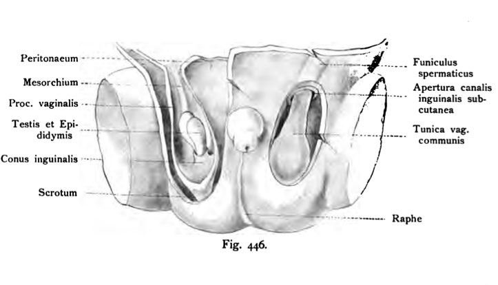
Fig. 446. Testicular descent
Human fetus of 25 cm head steißlänge. (Anatomical Collection in Basel)
The scrotum is opened on both sides. Right shows the tunica vaginalis communis with cremaster, somewhat isolated from the scrotum, Moreover, the inguinal canal, Apertura subcutanea visible.
On the left is the tunica vaginalis communis away, open the processus vaginalis of entire length, making testicles, epididymis, inguinal Conus, Mesorchium, as connective tissue between the processus vaginalis and the bottom of the Scrotum are visible.
- This text is a Google translate computer generated translation and may contain many errors.
Images from - Atlas of the Development of Man (Volume 2)
(Handatlas der entwicklungsgeschichte des menschen)
- Kollmann Atlas 2: Gastrointestinal | Respiratory | Urogenital | Cardiovascular | Neural | Integumentary | Smell | Vision | Hearing | Kollmann Atlas 1 | Kollmann Atlas 2 | Julius Kollmann
- Links: Julius Kollman | Atlas Vol.1 | Atlas Vol.2 | Embryology History
| Historic Disclaimer - information about historic embryology pages |
|---|
| Pages where the terms "Historic" (textbooks, papers, people, recommendations) appear on this site, and sections within pages where this disclaimer appears, indicate that the content and scientific understanding are specific to the time of publication. This means that while some scientific descriptions are still accurate, the terminology and interpretation of the developmental mechanisms reflect the understanding at the time of original publication and those of the preceding periods, these terms, interpretations and recommendations may not reflect our current scientific understanding. (More? Embryology History | Historic Embryology Papers) |
Reference
Kollmann JKE. Atlas of the Development of Man (Handatlas der entwicklungsgeschichte des menschen). (1907) Vol.1 and Vol. 2. Jena, Gustav Fischer. (1898).
Cite this page: Hill, M.A. (2024, April 24) Embryology Kollmann446.jpg. Retrieved from https://embryology.med.unsw.edu.au/embryology/index.php/File:Kollmann446.jpg
- © Dr Mark Hill 2024, UNSW Embryology ISBN: 978 0 7334 2609 4 - UNSW CRICOS Provider Code No. 00098G
Fig. 446. Descensus testicuiorum.
Menschlicher Fetus von 25 cm Kopf steißlänge. (Anatomische Sammlung in Basel)
Das Scrotum ist auf beiden Seiten geöffnet. Rechts zeigt sich die Tunica vaginalis communis mit dem Cremaster, etwas isoliert von dem Hodensack, überdies ist die Apertura canalis inguinalis subcutanea sichtbar. — Links ist die Tunica vaginalis communis entfernt, der Processus vaginalis geöffnet der ganzen Länge nach, wodurch Hoden, Nebenhoden, Conus inguinalis, Mesorchium, ebenso das Bindegewebe zwischen Processus vaginalis und dem Grund des Hodensackes erkennbar sind.
File history
Click on a date/time to view the file as it appeared at that time.
| Date/Time | Thumbnail | Dimensions | User | Comment | |
|---|---|---|---|---|---|
| current | 21:45, 16 October 2011 |  | 723 × 414 (38 KB) | S8600021 (talk | contribs) | {{Kollmann1907}} |
You cannot overwrite this file.
File usage
The following 3 pages use this file:

