File:Kollmann359.jpg

Original file (821 × 381 pixels, file size: 64 KB, MIME type: image/jpeg)
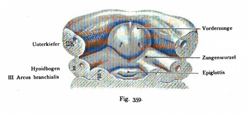
Fig. 359. Ventral wall of the foregut Human embryo of 12.5 mm
Ventral wall of the foregut with the gill arch, the inner gill-pouches, the investment of the tongue, front and behind the tongue, the latter = tongue root.
Nackensteißlänge.
The tuberculum impar has increased considerably, so that it already the impression of a tongue (its front section) makes immediately then the tongue root is easily recognizable, especially when compared with the next stage of development, the primitive root of tongue-summarizes the V-shaped front arising from the tuberculum impar tongue. by Together, the shift of the ventral ends of the gill arch, the furcula their shape changed considerably and has become a transverse column the anterior wall of the depressed epiglottis is formed.
- This text is a Google translate computer generated translation and may contain many errors.
Images from - Atlas of the Development of Man (Volume 2)
(Handatlas der entwicklungsgeschichte des menschen)
- Kollmann Atlas 2: Gastrointestinal | Respiratory | Urogenital | Cardiovascular | Neural | Integumentary | Smell | Vision | Hearing | Kollmann Atlas 1 | Kollmann Atlas 2 | Julius Kollmann
- Links: Julius Kollman | Atlas Vol.1 | Atlas Vol.2 | Embryology History
| Historic Disclaimer - information about historic embryology pages |
|---|
| Pages where the terms "Historic" (textbooks, papers, people, recommendations) appear on this site, and sections within pages where this disclaimer appears, indicate that the content and scientific understanding are specific to the time of publication. This means that while some scientific descriptions are still accurate, the terminology and interpretation of the developmental mechanisms reflect the understanding at the time of original publication and those of the preceding periods, these terms, interpretations and recommendations may not reflect our current scientific understanding. (More? Embryology History | Historic Embryology Papers) |
Reference
Kollmann JKE. Atlas of the Development of Man (Handatlas der entwicklungsgeschichte des menschen). (1907) Vol.1 and Vol. 2. Jena, Gustav Fischer. (1898).
Cite this page: Hill, M.A. (2024, April 24) Embryology Kollmann359.jpg. Retrieved from https://embryology.med.unsw.edu.au/embryology/index.php/File:Kollmann359.jpg
- © Dr Mark Hill 2024, UNSW Embryology ISBN: 978 0 7334 2609 4 - UNSW CRICOS Provider Code No. 00098G
Fig. 359. Ventrale Wand des Kopfdarms mit den Kiemenbogen, den inneren Kiementaschen, der Anlage der Zunge, Vorder- und Hinterzunge, die letztere = Zungenwurzel. Menschlicher Embryo von 12,5 mm
Nackensteißlänge.
Das Tuberculum impar hat sich beträchtlich vergrößert, so daß es schon den Eindruck einer Zunge — ihres vorderen Abschnittes — macht. Unmittelbar anschließend ist die Zungenwurzel schon zu erkennen, namentlich bei der Ver- ^leichung mit der folgenden Entwicklungsstufe; die primitive Zungenwurzel um- faßt V-förmig die aus dem Tuberculum impar entstandene Vorderzunge. Durch die Zusammenschiebung der ventralen Enden der Kiemenbogen hat die^Furcula ihre Form beträchtlich verändert und ist zu einer querliegenden Spalte geworden, deren vordere Wand von dem niedergedrückten Kehldeckel gebildet wird.
File history
Click on a date/time to view the file as it appeared at that time.
| Date/Time | Thumbnail | Dimensions | User | Comment | |
|---|---|---|---|---|---|
| current | 13:01, 16 October 2011 |  | 821 × 381 (64 KB) | S8600021 (talk | contribs) | {{Kollmann1907}} |
You cannot overwrite this file.
File usage
The following page uses this file:
