File:Kollmann347.jpg
Kollmann347.jpg (734 × 576 pixels, file size: 57 KB, MIME type: image/jpeg)
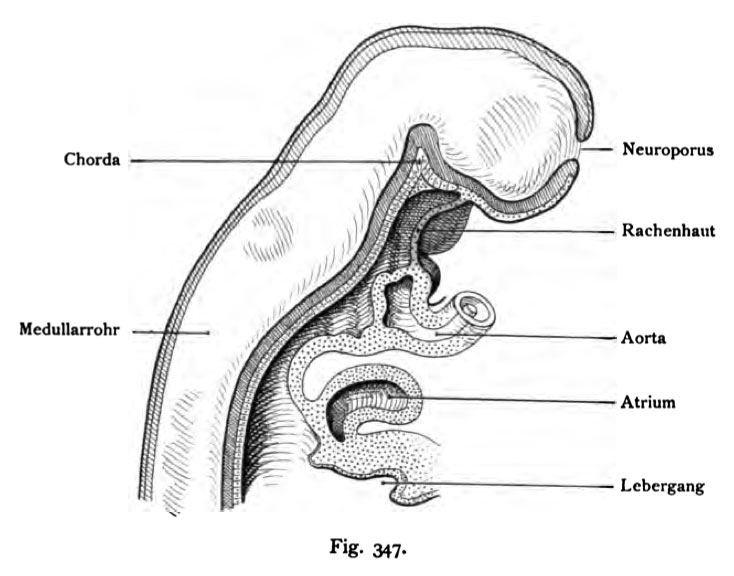
Fig. 347. The Pharyngeal Membrane of Human Embryo 3.1 mm CRL two weeks old
(After His.)
Revenge of the dorsal skin begins at the free end of the lower jaw protrusion moves posteriorly and slightly arched towards the base of the skull. Prior to this throat Skin is the bay mouth (red), behind the head is followed by the intestine. in the Bay mouth is ectoderm, endoderm in the foregut. Revenge of the Fallen skin consists of a layer of ectoderm and a layer of endoderm. the neural tube is at the front end is not closed: the anterior opening heifit Neuropore.
- This text is a Google translate computer generated translation and may contain many errors.
Images from - Atlas of the Development of Man (Volume 2)
(Handatlas der entwicklungsgeschichte des menschen)
- Kollmann Atlas 2: Gastrointestinal | Respiratory | Urogenital | Cardiovascular | Neural | Integumentary | Smell | Vision | Hearing | Kollmann Atlas 1 | Kollmann Atlas 2 | Julius Kollmann
- Links: Julius Kollman | Atlas Vol.1 | Atlas Vol.2 | Embryology History
| Historic Disclaimer - information about historic embryology pages |
|---|
| Pages where the terms "Historic" (textbooks, papers, people, recommendations) appear on this site, and sections within pages where this disclaimer appears, indicate that the content and scientific understanding are specific to the time of publication. This means that while some scientific descriptions are still accurate, the terminology and interpretation of the developmental mechanisms reflect the understanding at the time of original publication and those of the preceding periods, these terms, interpretations and recommendations may not reflect our current scientific understanding. (More? Embryology History | Historic Embryology Papers) |
Reference
Kollmann JKE. Atlas of the Development of Man (Handatlas der entwicklungsgeschichte des menschen). (1907) Vol.1 and Vol. 2. Jena, Gustav Fischer. (1898).
Cite this page: Hill, M.A. (2024, April 23) Embryology Kollmann347.jpg. Retrieved from https://embryology.med.unsw.edu.au/embryology/index.php/File:Kollmann347.jpg
- © Dr Mark Hill 2024, UNSW Embryology ISBN: 978 0 7334 2609 4 - UNSW CRICOS Provider Code No. 00098G
Fig. 347. Die Rachenhaut bei einem menschh*chen Embryo
3,1 mm Nackenlänge, zwei Wochen alt.
(Nach His.)
Die Rachenhaut beginnt am freien dorsalen Ende des Unterkieferfortsatzes und zieht leicht dorsal gewölbt nach der Basis des Schädels. Vor dieser Rachen- haut liegt die Mundbucht (rot), dahinter schließt sich der Kopfdarm an. In der Mundbucht befindet sich Ektoderm, im Kopfdarm Entoderm. Die Rachenhaut besteht aus einer Lage Ekto- und einer Lage Entoderm. Das Medullarrohr ist am vordersten Ende noch nicht geschlossen: die Öffnung heifit vorderer Neuroporus.
File history
Click on a date/time to view the file as it appeared at that time.
| Date/Time | Thumbnail | Dimensions | User | Comment | |
|---|---|---|---|---|---|
| current | 12:56, 16 October 2011 |  | 734 × 576 (57 KB) | S8600021 (talk | contribs) | {{Kollmann1907}} |
You cannot overwrite this file.
File usage
The following page uses this file:

