File:Kollmann083.jpg
From Embryology
Size of this preview: 474 × 600 pixels. Other resolution: 632 × 800 pixels.
Original file (632 × 800 pixels, file size: 41 KB, MIME type: image/jpeg)
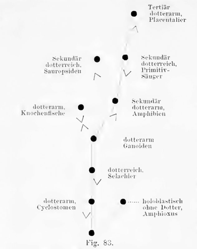
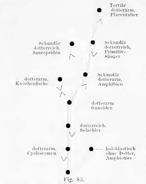
Fig. 83. The varying amounts of yolk in the eggs of vertebrates
as shown schematically.
after Rabl.
- This text is a Google translate computer generated translation and may contain many errors.
Images from - Atlas of the Development of Man (Volume 1)
(Handatlas der entwicklungsgeschichte des menschen)
- Kollmann Atlas 1: Predevelopment | Ontogeny | Fetal membranes | Body shape | Systems and organs | Kollmann Atlas 1 | Kollmann Atlas 2 | Julius Kollmann
- Links: Julius Kollman | Atlas Vol.1 | Atlas Vol.2 | Embryology History
| Historic Disclaimer - information about historic embryology pages |
|---|
 |
Fig. 83. Die wechselnden Mengi-n des Dotters in den Eiern der Wirbeltiere, schematisch dargestellt. Nach Rabl.
File history
Click on a date/time to view the file as it appeared at that time.
| Date/Time | Thumbnail | Dimensions | User | Comment | |
|---|---|---|---|---|---|
| current | 16:05, 30 October 2011 | | 632 × 800 (41 KB) | S8600021 (talk | contribs) | ==Fig. 83. == {{Kollmann1906}} Category:Mesoderm |
You cannot overwrite this file.
File usage
The following page uses this file: