File:Kingsbury1922 fig03.jpg

Original file (1,000 × 784 pixels, file size: 96 KB, MIME type: image/jpeg)
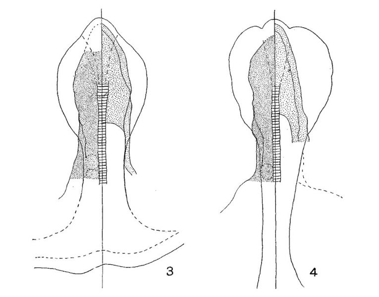
Figs. 3 and 4
Plottings from series cut in the transverse plane of Squalus acanthias embryos, 2.2 mm. and 2.6 mm. length, 6 and 8 somites, respectively.
These are the embryos of figures 7 and 9, plate 1.
In each, the notochord is indicated by cross-barring; the stipple areas on the right and left mark the extent of the entoderm and mesoderm, respectively; the two lines on the right bound the lateral wall of the archenteric cavity; the outline of the median ‘wedge—shaped area’ is shown in broken line, and closely corresponds to the territory of the prechordal plate at these stages.
Reference
Kingsbury BF. The fundamental plan of the vertebrate brain. (1922) J. Comp. Neural. 461-490.
Cite this page: Hill, M.A. (2024, April 19) Embryology Kingsbury1922 fig03.jpg. Retrieved from https://embryology.med.unsw.edu.au/embryology/index.php/File:Kingsbury1922_fig03.jpg
- © Dr Mark Hill 2024, UNSW Embryology ISBN: 978 0 7334 2609 4 - UNSW CRICOS Provider Code No. 00098G
File history
Click on a date/time to view the file as it appeared at that time.
| Date/Time | Thumbnail | Dimensions | User | Comment | |
|---|---|---|---|---|---|
| current | 10:05, 23 November 2019 |  | 1,000 × 784 (96 KB) | Z8600021 (talk | contribs) | |
| 10:03, 23 November 2019 |  | 1,371 × 1,449 (325 KB) | Z8600021 (talk | contribs) |
You cannot overwrite this file.
File usage
The following page uses this file: