File:Keith1921 fig046.jpg
Original file (956 × 1,081 pixels, file size: 79 KB, MIME type: image/jpeg)
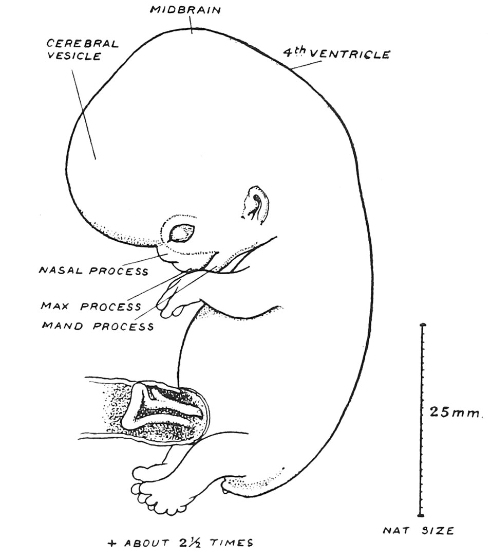
Fig. 46. Outline of a Foetus 22 mm long
And at the end of the 2nd month of development. (After Broman.)
At the end of the 8th week the crownrump diameter measures about 25 mm. (1 inch). The changes of this week are a continuation of those we have just described (Fig. 46) ; the nasal and maxillary processes have fused to form the upper face ; the upper lip is completed, but the palatal processes have not yet separated the buccal from the nasal cavities. The cerebral vesicles are expanding rapidly backwards ; the neck is being differentiated and the limbs are making progress. The rudiment of the external genital organs is apparent, but as yet gives no clue to sex. The intestinal loop lies within the root of the umbilical cord. Henceforward, until the end of gestation, the chief changes are those of growth.[1]
- Chapter 4 Figures: 42 | 43 | 44 | 45 | 46 | All Figures
| Historic Disclaimer - information about historic embryology pages |
|---|
 |
Reference
Keith A. Human Embryology and Morphology. (1921) New York, Longmans, Green & Co. London: Edward Arnold.
Human Embryology and Morphology: 1 Early Ovum and Embryo | 2 Connection between Foetus and Uterus | 3 Primitive Streak Notochord and Somites | 4 Age Changes | 5 Spinal Column and Back | 6 Body Segmentation | 7 Spinal Cord | 8 Mid- and Hind-Brains | 9 Fore-Brain | 10 Fore-Brain Cerebral Vesicles | 11 Cranium | 12 Face | 13 Teeth and Mastication | 14 Nasal and Olfactory | 15 Sense OF Sight | 16 Hearing | 17 Pharynx and Neck | 18 Tongue, Thyroid and Pharynx | 19 Organs of Digestion | 20 Circulatory System | 21 Circulatory System (continued) | 22 Respiratory System | 23 Urogenital System | 24 Urogenital System (Continued) | 25 Body Wall and Pelvic Floor | 26 Limb Buds | 27 Limbs | 28 Skin and Appendages | Figures
Cite this page: Hill, M.A. (2024, April 23) Embryology Keith1921 fig046.jpg. Retrieved from https://embryology.med.unsw.edu.au/embryology/index.php/File:Keith1921_fig046.jpg
- © Dr Mark Hill 2024, UNSW Embryology ISBN: 978 0 7334 2609 4 - UNSW CRICOS Provider Code No. 00098G
- ↑ For changes in 9th week see F. E. Blaisdell, Journ. Anat. 1914, vol. 48, p. 182.
File history
Click on a date/time to view the file as it appeared at that time.
| Date/Time | Thumbnail | Dimensions | User | Comment | |
|---|---|---|---|---|---|
| current | 12:41, 23 December 2014 | | 956 × 1,081 (79 KB) | Z8600021 (talk | contribs) | |
| 12:37, 23 December 2014 |  | 1,200 × 1,175 (103 KB) | Z8600021 (talk | contribs) |
You cannot overwrite this file.
File usage
The following 3 pages use this file: