File:Keith1921 fig035.jpg
Original file (984 × 808 pixels, file size: 143 KB, MIME type: image/jpeg)
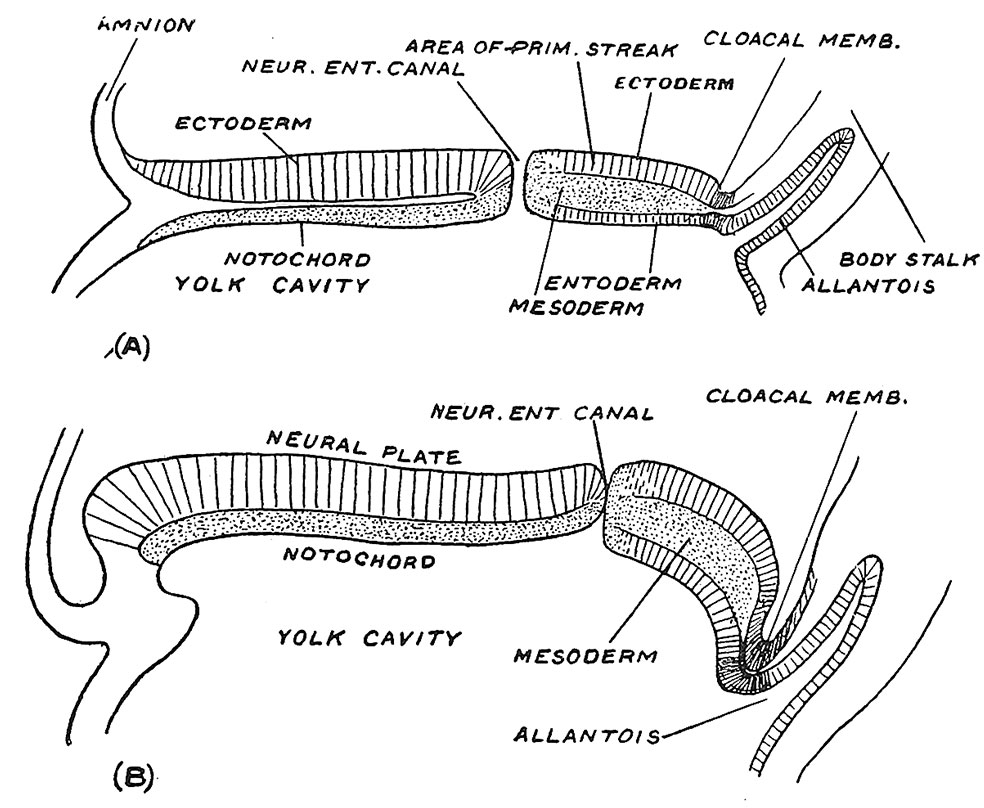
Fig. 35. Sections along the median line of two embryonic plates
Figured by Graf Spee, to show the shifting backwards of the neurenteric canal and primitive streak as growth takes place.
If sections are made along the embryonic plate (Fig. 35, A and B) further light is thrown on the relationship of the neurenteric canal and primitive streak to the growth of the embryo. In Fig. 35, A the neurenteric canal is seen to be placed near the middle point of the plate — which has a total length of a little over 1 mm. while in the older embryo which measures r7 mm. in length, it is pushed backwards by the rapid growth and extension of the j^recanalicular part of the embryonic plate. The region of the primitive streak — the postcanalicular part of the embryonic plate — although the site of mesodermal production, has undergone a lesser degree of growth and is being pushed to the hinder end of the embryonic plate. The exact manner in which the precanalicular part expands we are not certain of, but it will be noted that at the anterior lip of the neurenteric canal the neural plate becomes continuous with the notochordal plate and we suspect that this lip represents a growing edge. The first formed part of the precanalicular plate represents the hinder cranial region ; as the plate grows the neurenteric canal moves backwards through the cervical and dorsal regions until it reaches the lumbar region early in the fourth week, the embryo then being less than 3 mm. in length. If, as sometimes occurs, the neurenteric canal remains unclosed, a fistula from the bowel opens on the lumbar region of the back. In Fig. 34 it will be seen that while the neurenteric canal lies at the anterior end of the primitive streak a very important structure — the cloacal membrane — marks its posterior end. The cloacal membrane, lying at the foot of the body stalk, marks the site of the anus and vulval cleft. Thus the whole of the hinder end of the human body is developed on each side of the primitive streak, a relationship which must be understood if certain malformations of the human body are to be adequately explained.
- Chapter 3 Figures: 34 | 35 | 36 | 37 | 38 | 39 | 40 | 41 | All Figures
| Historic Disclaimer - information about historic embryology pages |
|---|
 |
Reference
Keith, A. Human Embryology And Morphology (1921) Longmans, Green & Co.:New York.
Human Embryology and Morphology: 1 Early Ovum and Embryo | 2 Connection between Foetus and Uterus | 3 Primitive Streak Notochord and Somites | 4 Age Changes | 5 Spinal Column and Back | 6 Body Segmentation | 7 Spinal Cord | 8 Mid- and Hind-Brains | 9 Fore-Brain | 10 Fore-Brain Cerebral Vesicles | 11 Cranium | 12 Face | 13 Teeth and Mastication | 14 Nasal and Olfactory | 15 Sense OF Sight | 16 Hearing | 17 Pharynx and Neck | 18 Tongue, Thyroid and Pharynx | 19 Organs of Digestion | 20 Circulatory System | 21 Circulatory System (continued) | 22 Respiratory System | 23 Urogenital System | 24 Urogenital System (Continued) | 25 Body Wall and Pelvic Floor | 26 Limb Buds | 27 Limbs | 28 Skin and Appendages | Figures
Cite this page: Hill, M.A. (2024, April 16) Embryology Keith1921 fig035.jpg. Retrieved from https://embryology.med.unsw.edu.au/embryology/index.php/File:Keith1921_fig035.jpg
- © Dr Mark Hill 2024, UNSW Embryology ISBN: 978 0 7334 2609 4 - UNSW CRICOS Provider Code No. 00098G
File history
Click on a date/time to view the file as it appeared at that time.
| Date/Time | Thumbnail | Dimensions | User | Comment | |
|---|---|---|---|---|---|
| current | 11:38, 23 December 2014 | | 984 × 808 (143 KB) | Z8600021 (talk | contribs) | |
| 11:33, 23 December 2014 | 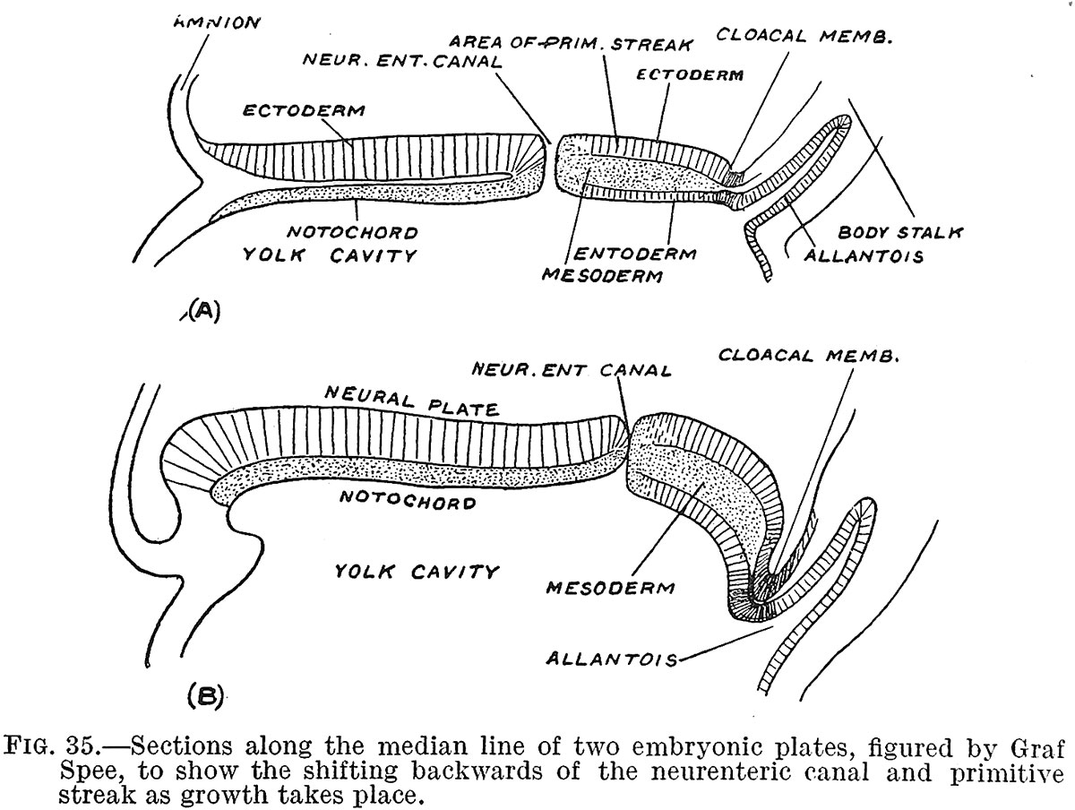 | 1,200 × 904 (181 KB) | Z8600021 (talk | contribs) |
You cannot overwrite this file.
File usage
The following 2 pages use this file: