File:Keibel Mall 2 624.jpg
Original file (800 × 725 pixels, file size: 93 KB, MIME type: image/jpeg)
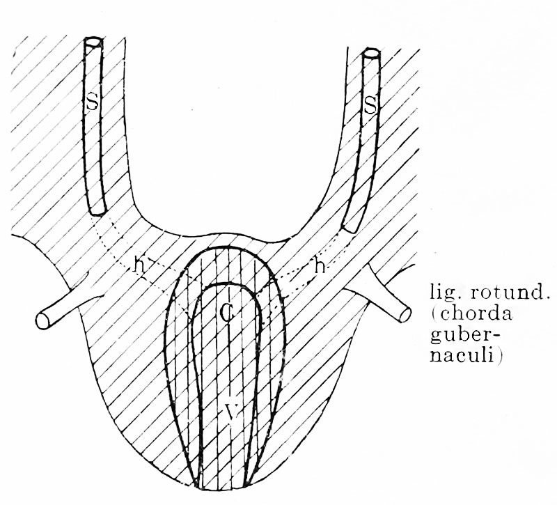
Fig. 624. Diagram of the development of the male uterus
Only the utero-vaginal canal becomes enclosed by the mesenchymatous uterine wall, the horizontal portions of both primitive tubes had previously degenerated. Consequently the plica inguinalis, or the chorda gubernaculi which is derived from it, does not come into relation with the uterus.
| Embryology - 20 Apr 2024 |
|---|
| Google Translate - select your language from the list shown below (this will open a new external page) |
|
العربية | català | 中文 | 中國傳統的 | français | Deutsche | עִברִית | हिंदी | bahasa Indonesia | italiano | 日本語 | 한국어 | မြန်မာ | Pilipino | Polskie | português | ਪੰਜਾਬੀ ਦੇ | Română | русский | Español | Swahili | Svensk | ไทย | Türkçe | اردو | ייִדיש | Tiếng Việt These external translations are automated and may not be accurate. (More? About Translations) |
Felix W. The development of the urinogenital organs. In Keibel F. and Mall FP. Manual of Human Embryology II. (1912) J. B. Lippincott Company, Philadelphia. pp 752-979.
| Historic Disclaimer - information about historic embryology pages |
|---|
 |
Cite this page: Hill, M.A. (2024, April 20) Embryology Keibel Mall 2 624.jpg. Retrieved from https://embryology.med.unsw.edu.au/embryology/index.php/File:Keibel_Mall_2_624.jpg
- © Dr Mark Hill 2024, UNSW Embryology ISBN: 978 0 7334 2609 4 - UNSW CRICOS Provider Code No. 00098G
File history
Click on a date/time to view the file as it appeared at that time.
| Date/Time | Thumbnail | Dimensions | User | Comment | |
|---|---|---|---|---|---|
| current | 11:11, 12 November 2018 | | 800 × 725 (93 KB) | Z8600021 (talk | contribs) | |
| 11:08, 12 November 2018 |  | 928 × 1,169 (162 KB) | Z8600021 (talk | contribs) | Fig. 624. Diagram of the development of the male uterus. Only the utero-vaginal canal becomes enclosed by the mesenchymatous uterine wall, the horizontal portions of both primitive tubes had previously degenerated. Consequently the plica inguinalis, or... |
You cannot overwrite this file.
File usage
The following 2 pages use this file: