File:Keibel Mall 2 623.jpg

Original file (1,549 × 1,709 pixels, file size: 437 KB, MIME type: image/jpeg)
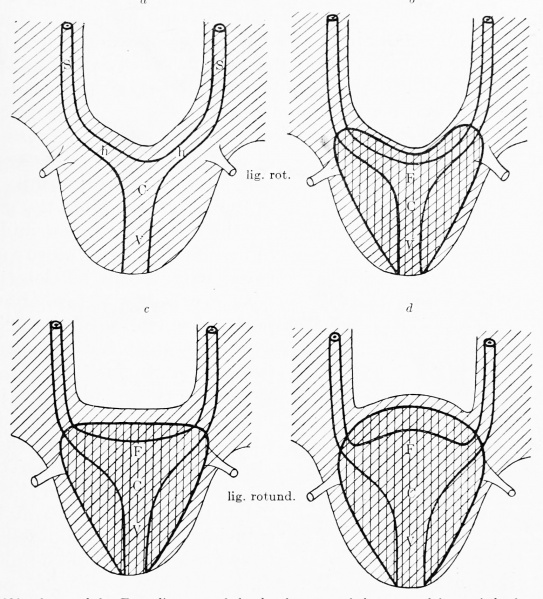
Fig. 623 a, b, c, and d. Four diagrams of the development of the external form of the female uterus
The mesonephric folds and the genital cord are shaded obliquely, the mesenchymatous uterine wall vertically. The various parts of the primitive uterine wall and of the utero-vaginal canal are lettered. S vertical portion; h, horizontal portion of the wall of the primitive tube; F, fundus uteri; C, cervix uteri; V, vagina.
Diagram a shows the position of the primitive tubes and of the utero-vaginal canal after it is completed.
Diagram b shows the relation of the mesenchymatous uterine wall to the primitive tubes and to the utero-vaginal canal. It encloses the whole of the horizontal portion of both tubes in the uterine region and as a result the lig. rotundum is brought into relation with the uterine wall. The fundus uteri is bent in at an angle (uterus introrsum arcuatus).
Diagram c shows the broadening of the horizontal portion of both tubes to form the fundus uteri, the broadening taking place in such a way as to straighten out the inward bend {uterus plani/undus) .
Diagram d The broadening of the fundus has increased and it is curved outward (uterus foras arcuatus). lig. rotund, (chorda gubernaculi)
| Embryology - 18 Apr 2024 |
|---|
| Google Translate - select your language from the list shown below (this will open a new external page) |
|
العربية | català | 中文 | 中國傳統的 | français | Deutsche | עִברִית | हिंदी | bahasa Indonesia | italiano | 日本語 | 한국어 | မြန်မာ | Pilipino | Polskie | português | ਪੰਜਾਬੀ ਦੇ | Română | русский | Español | Swahili | Svensk | ไทย | Türkçe | اردو | ייִדיש | Tiếng Việt These external translations are automated and may not be accurate. (More? About Translations) |
Felix W. The development of the urinogenital organs. In Keibel F. and Mall FP. Manual of Human Embryology II. (1912) J. B. Lippincott Company, Philadelphia. pp 752-979.
| Historic Disclaimer - information about historic embryology pages |
|---|
 |
Cite this page: Hill, M.A. (2024, April 18) Embryology Keibel Mall 2 623.jpg. Retrieved from https://embryology.med.unsw.edu.au/embryology/index.php/File:Keibel_Mall_2_623.jpg
- © Dr Mark Hill 2024, UNSW Embryology ISBN: 978 0 7334 2609 4 - UNSW CRICOS Provider Code No. 00098G
File history
Click on a date/time to view the file as it appeared at that time.
| Date/Time | Thumbnail | Dimensions | User | Comment | |
|---|---|---|---|---|---|
| current | 11:17, 12 November 2018 | | 1,549 × 1,709 (437 KB) | Z8600021 (talk | contribs) | |
| 11:14, 12 November 2018 | 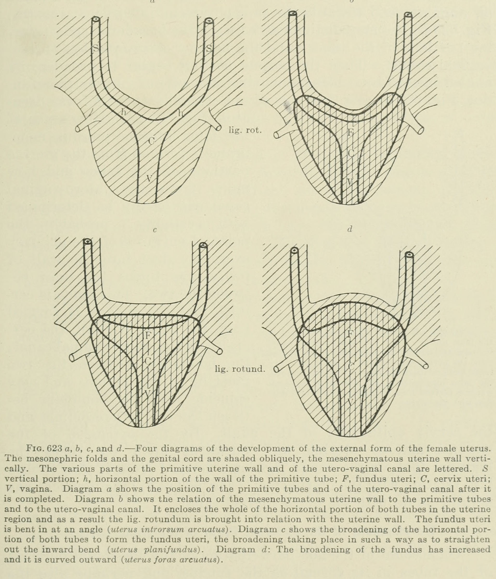 | 1,907 × 2,223 (562 KB) | Z8600021 (talk | contribs) | Fig. 623 a, b, c, and d. Four diagrams of the development of the external form of the female uterus. The mesonephric folds and the genital cord are shaded obliquely, the mesenchymatous uterine wall vertically. The various parts of the primitive uterin... |
You cannot overwrite this file.
File usage
The following 2 pages use this file: