File:Keibel Mall 2 573.jpg
Original file (1,280 × 886 pixels, file size: 165 KB, MIME type: image/jpeg)
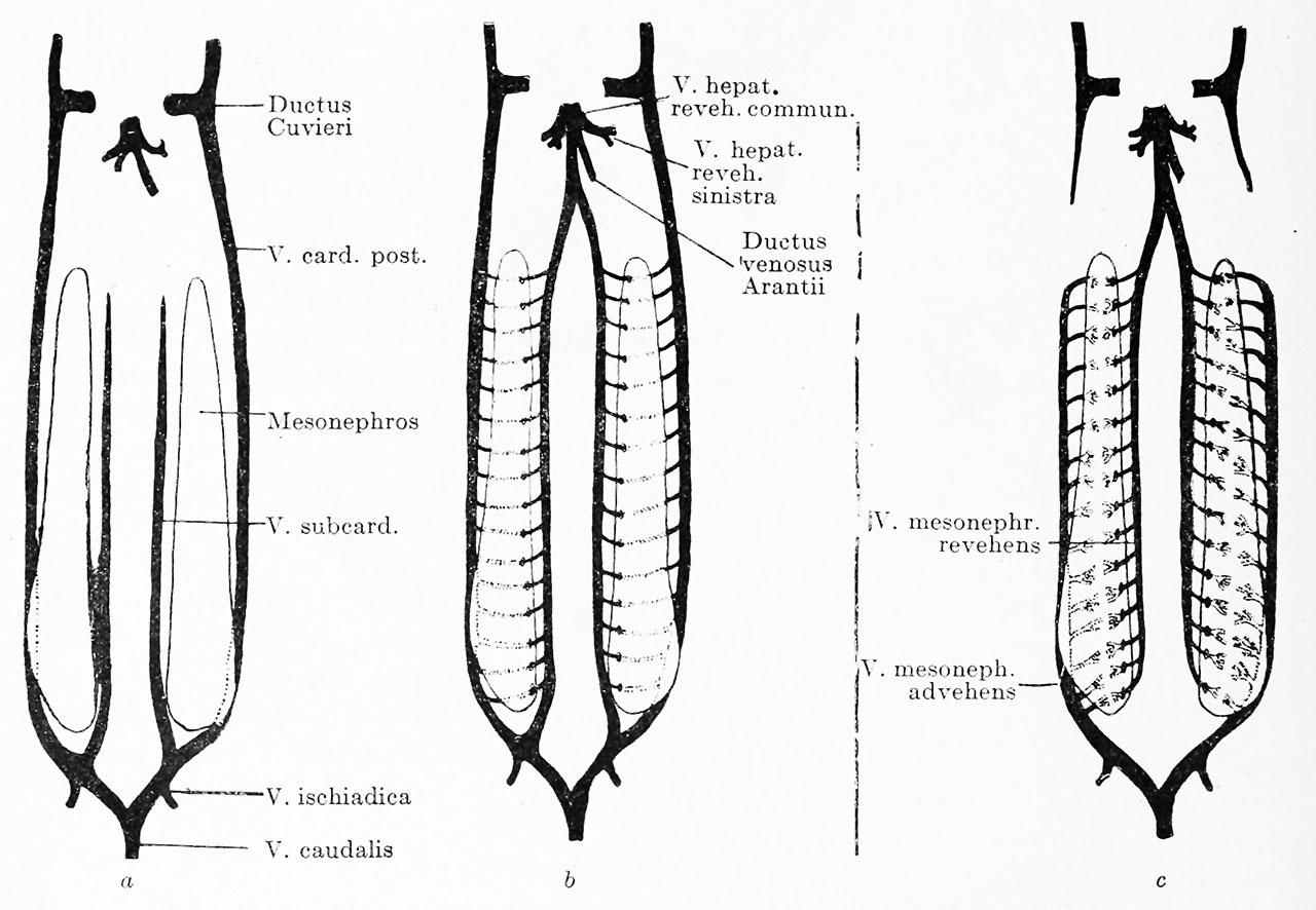
Fig. 573 a, b and c. Plan of the development of the mesonephric portal circulation
| a. A vein runs on both sides of the mesonephros, the posterior cardinal laterally and dorsally and the subcardinal medially and ventrally. The posterior cardinal opens cranially into the ductus Cuvieri, while the subcardinal ends blindly. Caudally both veins are connected with the paired v. ischiadica and the unpaired v. caudalis. | b. Transverse anastomoses have formed between the posterior cardinal and the subcardinal, traversing the mesonephros. The subcardinal opens into the v. hepatica revehens communis, | c. The cardinal has lost its connection with the ductus Cuvieri, but retains that with the vv. ischiadica and caudalis. The subcardinal has retained its opening into the v. hepatica revehens communis, but has lost its connection with the vv. ischiadica and caudalis. Capillary networks have developed in the transverse anastomoses. The blood of the vv. caudalis and ischiadica must now take the following course: 1, v. cardinalis, 2, the transverse anastomoses with capillary networks inside the mesonephros, 3, v. subcardinalis, 4, v. hepatica revehens communis, 5, sinus venosus. |
| Embryology - 19 Apr 2024 |
|---|
| Google Translate - select your language from the list shown below (this will open a new external page) |
|
العربية | català | 中文 | 中國傳統的 | français | Deutsche | עִברִית | हिंदी | bahasa Indonesia | italiano | 日本語 | 한국어 | မြန်မာ | Pilipino | Polskie | português | ਪੰਜਾਬੀ ਦੇ | Română | русский | Español | Swahili | Svensk | ไทย | Türkçe | اردو | ייִדיש | Tiếng Việt These external translations are automated and may not be accurate. (More? About Translations) |
Felix W. The development of the urinogenital organs. In Keibel F. and Mall FP. Manual of Human Embryology II. (1912) J. B. Lippincott Company, Philadelphia. pp 752-979.
| Historic Disclaimer - information about historic embryology pages |
|---|
 |
Cite this page: Hill, M.A. (2024, April 19) Embryology Keibel Mall 2 573.jpg. Retrieved from https://embryology.med.unsw.edu.au/embryology/index.php/File:Keibel_Mall_2_573.jpg
- © Dr Mark Hill 2024, UNSW Embryology ISBN: 978 0 7334 2609 4 - UNSW CRICOS Provider Code No. 00098G
File history
Click on a date/time to view the file as it appeared at that time.
| Date/Time | Thumbnail | Dimensions | User | Comment | |
|---|---|---|---|---|---|
| current | 22:07, 13 November 2018 | | 1,280 × 886 (165 KB) | Z8600021 (talk | contribs) | |
| 22:06, 13 November 2018 | 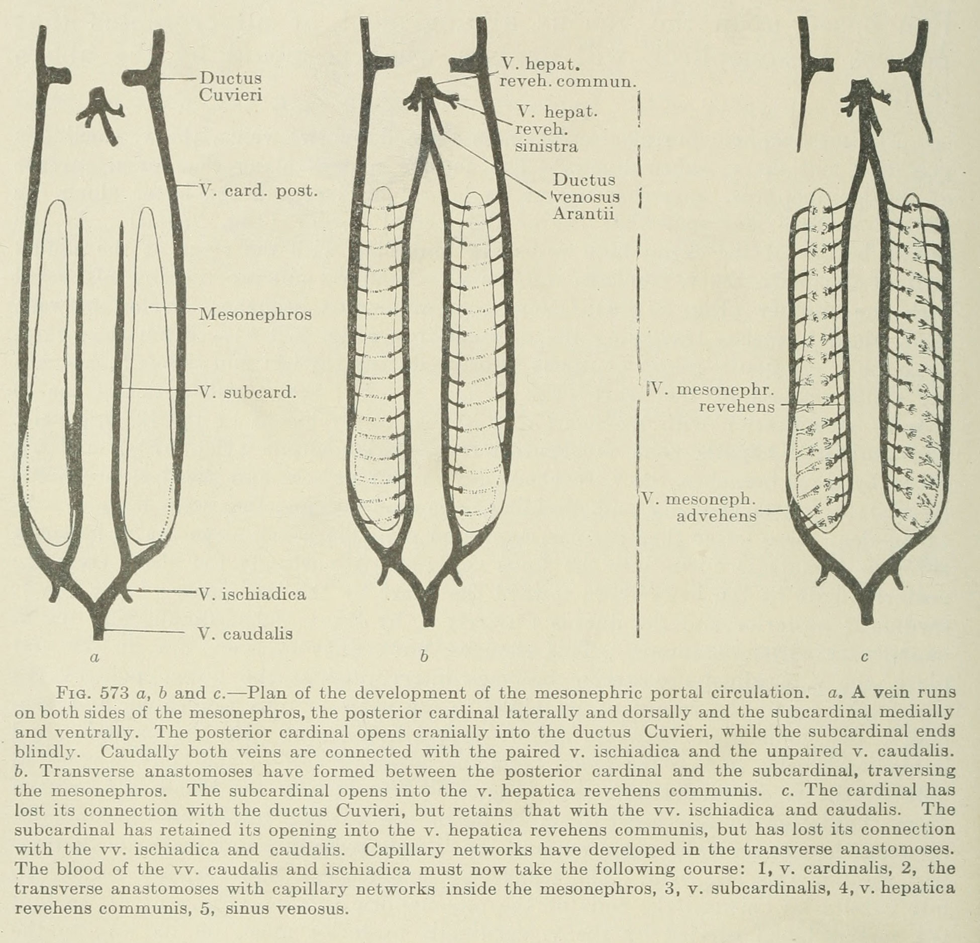 | 1,950 × 1,876 (486 KB) | Z8600021 (talk | contribs) | Fig. 573 a, b and c. Plan of the development of the mesonephric portal circulation, a. A vein runs on both sides of the mesonephros, the posterior cardinal laterally and dorsally and the subcardinal medially and ventrally. The posterior cardinal opens... |
You cannot overwrite this file.
File usage
The following 2 pages use this file: