File:Keibel Mall 2 493.jpg
Original file (680 × 1,000 pixels, file size: 89 KB, MIME type: image/jpeg)
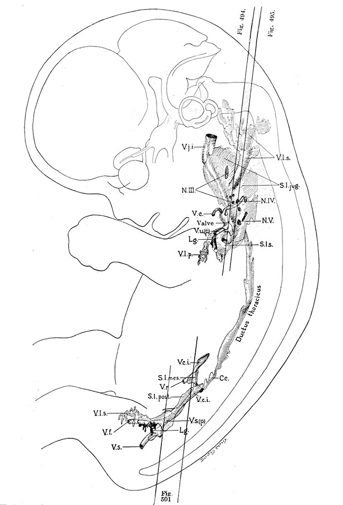
Fig. 493. Profile reconstruction of the primitive lymphatic system in a human embryo 30 mm long
Mall's collection, No. 86. x about 5.8
C.c, cisterna chyli; L.g., lymphoglandula; N.III., N.IV., and if. V., nervi cervicalis; S.l.jug., saccus lynphaticus jugularis; S.l.mes., saccus lymphaticus retroperitonalis; S.l.p., saccus lymphaticus posterior; S.l.s., saccus lymphaticus subclavius; Y.c, vena cephalica; V.e.i., vena cava inferior; Y.f., vena femoralis; V.j.i., vena jugularis interna; Y.l.p., vasa lymphatica profunda; Y.l.s., vasa lymphatica superficialia; Y r., vena renalis; Y.s., vena sciatica; V.u.(p.), vena ulnaris (primitiva I.
- IV. The Development of the Lymphatic System: Chapter XVIII. Development of Blood, Vascular System, and Spleen | Historic Disclaimer
Reference
Sabin FR. The Development of the Lymphatic System in Keibel F. and Mall FP. Manual of Human Embryology II. (1912) J. B. Lippincott Company, Philadelphia.
Cite this page: Hill, M.A. (2024, April 18) Embryology Keibel Mall 2 493.jpg. Retrieved from https://embryology.med.unsw.edu.au/embryology/index.php/File:Keibel_Mall_2_493.jpg
- © Dr Mark Hill 2024, UNSW Embryology ISBN: 978 0 7334 2609 4 - UNSW CRICOS Provider Code No. 00098G
File history
Click on a date/time to view the file as it appeared at that time.
| Date/Time | Thumbnail | Dimensions | User | Comment | |
|---|---|---|---|---|---|
| current | 14:29, 2 March 2014 | | 680 × 1,000 (89 KB) | Z8600021 (talk | contribs) |
You cannot overwrite this file.
File usage
The following 2 pages use this file: