File:Keibel Mall 2 032.jpg

Original file (1,200 × 750 pixels, file size: 139 KB, MIME type: image/jpeg)
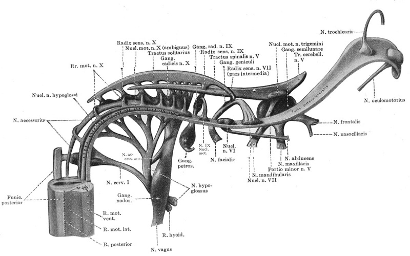
Fig. 32. Reconstruction showing the cranial nerves in a 10 mm human embryo
(Huber collection No. 3).
The brain wall is removed so as to show the primary sensory paths and motor nuclei of the different nerves.
The sensory fibres enter the marginal zone near the junction of the basal and alar plates, and immediately form longitudinal tracts analogous to the posterior funiculi of the spinal cord. The entering fibres of the seventh, ninth and tenth nerves in this manner unite to form the tractus solitarius, as shown in Fig. 32 and Fig. 96. The entering fibres of the trigeminal nerve form a similar but separate bundle. In the latter case we can recognize a cephalic limb extending to the anlage of the cerebellum and midbrain and a caudal limb extending toward the spinal region. A similar but smaller tract is formed by the entering fibres of the acoustic nerve, which at this time consists mostly of vestibular fibres. In addition to the tracts mentioned there are present in the marginal zone a few early representatives of the correlating fibres which later form the formatio reticularis and system of arcuate fibres and their longitudinal extensions. Near the median line the marginal zone is somewhat thicker from the presence of such fibres.
| Historic Disclaimer - information about historic embryology pages |
|---|
 |
CNS Figures: 22 - neural-plate stage | 23 - 5 to 14 somites stage | 24 - 3.2 mm embryo | 30 - brain 3, 4 and 8 weeks | 32 - 10 mm cranial nerves
- XIV. The Development of the Nervous System: The Histogenesis of Nervous Tissue | The Development of the Central Nervous System | The Peripheral Nervous System | The Sympathetic Nervous System | Manual of Human Embryology II
Cite this page: Hill, M.A. (2024, April 25) Embryology Keibel Mall 2 032.jpg. Retrieved from https://embryology.med.unsw.edu.au/embryology/index.php/File:Keibel_Mall_2_032.jpg
- © Dr Mark Hill 2024, UNSW Embryology ISBN: 978 0 7334 2609 4 - UNSW CRICOS Provider Code No. 00098G
File history
Click on a date/time to view the file as it appeared at that time.
| Date/Time | Thumbnail | Dimensions | User | Comment | |
|---|---|---|---|---|---|
| current | 11:57, 24 January 2014 | | 1,200 × 750 (139 KB) | Z8600021 (talk | contribs) | ==Fig. 31. Reconstruction showing the cranial nerves in a 10 mm human embryo== (Huber collection No. 3). The brain wall is removed so as to show the primary sensory paths and motor nuclei of the different nerves. |
You cannot overwrite this file.
File usage
The following 3 pages use this file: